
Fig. S1
- ID
- ZDB-IMAGE-150416-29
- Genes
- Publication
- Wen et al., 2015 - Sox4 regulates choroid fissure closure by limiting hedgehog signaling during ocular morphogenesis
- All Figures
- Figures for Wen et al., 2015
|
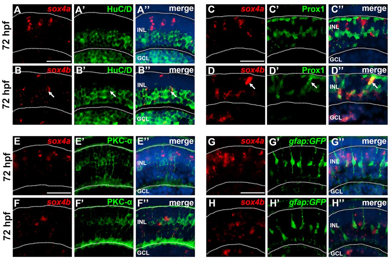
Fig. S1 Co-localization of sox4a/b with cell specific markers at 72 hpf. (A-B′′) FISH for sox4a and sox4b followed by immunostaining for the ganglion and amacrine cell marker HuC/D. Whereas sox4a did not co-localize with HuC/D (A-A′′), some sox4b-positive cells co-localized with HuC/D (B-B′′, arrows). (C-D′′) FISH for sox4a and sox4b followed by immunostaining for the horizontal cell marker Prox1. Sox4a did not co-localize with Prox1 (C-C′′); however some sox4b-positive cells in the outer half of the INL co-localized with Prox1 (D-D′′, arrows). (E-F′′) FISH for sox4a and sox4b followed by immunostaining for the bipolar cell marker PKC-α. No co-localization with PKC-α was detected. (G-H′′) FISH for sox4a and sox4b was performed in embryos transgenic for gfap:GFP which labels Müller glial cells. No co-localization with Müller cells was observed. All scale bars equal 25 µm. GCL, ganglion cell layer; INL, inner nuclear layer.
Reprinted from Developmental Biology, 399(1), Wen, W., Pillai-Kastoori, L., Wilson, S.G., Morris, A.C., Sox4 regulates choroid fissure closure by limiting hedgehog signaling during ocular morphogenesis, 139-53, Copyright (2015) with permission from Elsevier. Full text @ Dev. Biol.