
|
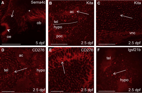
Fig. 3
Antibodies detect neural receptor proteins within discrete subcellular localisations. (A) Anti-Sema4c staining in the anterior neuroepithelium. (B) Anti-Kita labels the anterior (ac) and postoptic (poc) commissures and is also enriched at the periphery of cells (arrow). (C) Anti-Kita also stained axons of the ventral nerve cord (vnc) and the surface of hindbrain cells. Anti-CD276 staining in the developing midbrain (D), and tectum (E). (F) Anti-Igsf21b stained axon-rich regions in the anterior forebrain where it was often concentrated in discrete puncta (arrow). Views are anterior to the left and dorsal for A and E, lateral for B, C and F, ventral for D. Scale bars represent 100 µm in (A) and 50 µm in (B–F).