
Fig. 1
- ID
- ZDB-IMAGE-150224-1
- Genes
- Antibodies
- Publication
- Berger et al., 2014 - Loss of tropomodulin4 in the zebrafish mutant träge causes cytoplasmic rod formation and muscle weakness reminiscent of nemaline myopathy
- All Figures
- Figures for Berger et al., 2014
|
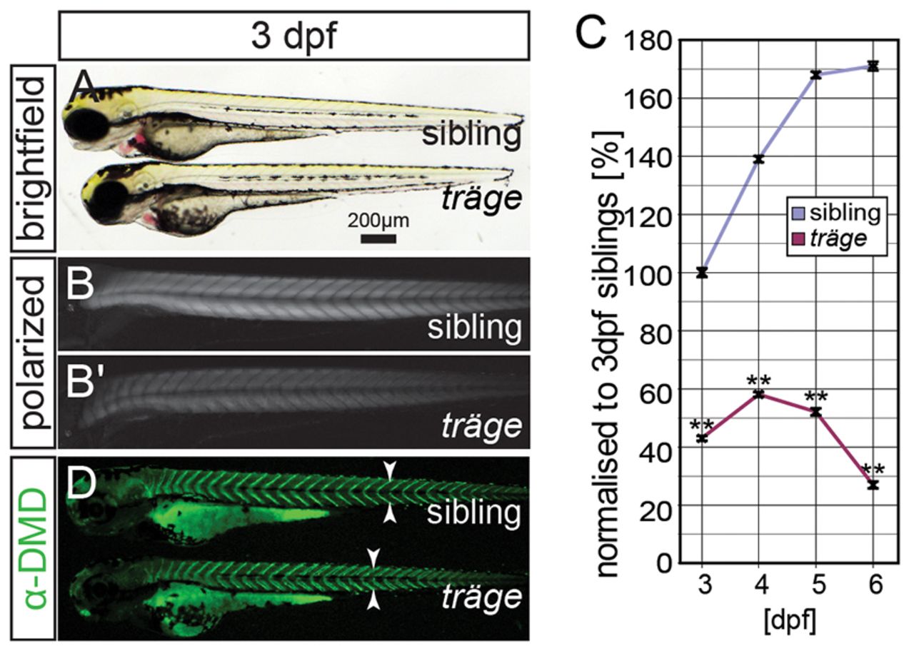
Fig. 1 The mutant träge (trg) shows a reduction in birefringence, indicating muscle damage. (A) Under brightfield microscopy, trg mutants appear similar to their wild-type siblings. (B) Under polarised light, the muscle of siblings appears brighter than that of the (B′) trg mutants due to a reduction in birefringence. (C) Quantification of the birefringence followed by normalization to that of 3-dpf-old siblings reveals that the birefringence of the siblings increases from 3 dpf to 6 dpf, roughly following a sigmoidal curve. By contrast, 3-dpf- to 6-dpf-old trg larvae show a highly significant reduction in birefringence when compared with that of 3-dpf siblings (P<0.01, n=3). (D) Immunohistochemistry with antibodies against dystrophin shows that dystrophin expression at the vertical myosepta (arrowheads) is unaffected in trg mutants. Data are means ± s.e.m., **P<0.01. Scale bar: 200 μm.