
Fig. 2
- ID
- ZDB-IMAGE-141007-235
- Genes
- Publication
- Cardozo et al., 2014 - Cdon acts as a Hedgehog decoy receptor during proximal-distal patterning of the optic vesicle
- All Figures
- Figures for Cardozo et al., 2014
|
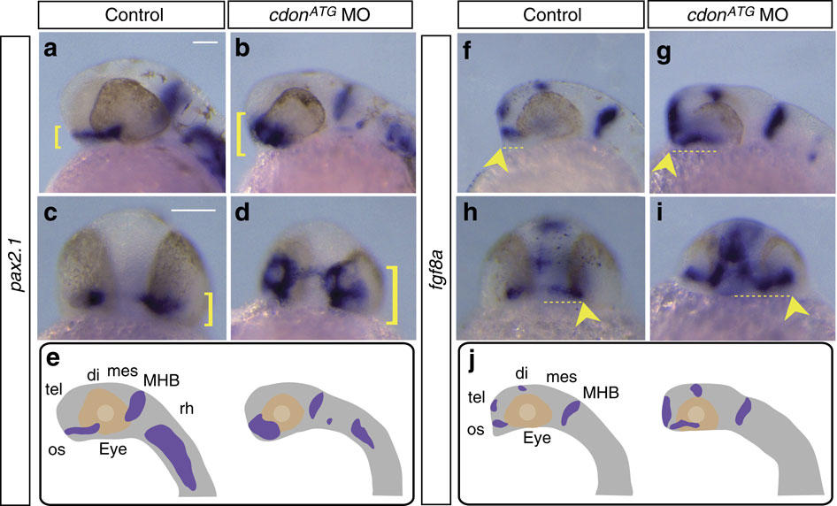
Fig. 2
(a–j) In situ hybridization analysis for two optic stalk markers, pax2.1 (a–d) and fgf8a (f–i) at 28 hpf and 24 hpf, respectively. Embryos are shown in lateral (a,b,f,g) and frontal (c,d,h,i) views. Expression patterns of both genes are schematically represented in e and j. In cdonATG morphants, pax2.1 expression is expanded dorsally in the optic stalk (b,d brackets) when compared with controls (a,c brackets). Fgf8a expression is expanded caudally and laterally in the optic stalk (g,i arrowhead and dotted lines) as well as in the telencephalon (g,i) when compared with control embryos (f,h arrowhead and dotted lines). di, diencephalon; mes, mesencephalon; MHB, midbrain–hindbrain boundary; os, optic stalk; rh, rhombencephalon; tel, telencephalon. Scale bars, 100 μm.