
Fig. 8
- ID
- ZDB-IMAGE-140915-12
- Publication
- Skobo et al., 2014 - Zebrafish ambra1a and ambra1b Knockdown Impairs Skeletal Muscle Development
- All Figures
- Figures for Skobo et al., 2014
|
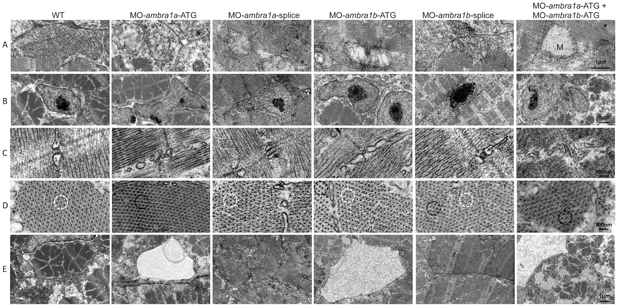
Fig. 8
Ultrastructural analysis of ambra1 morphants muscles reveals disorganized subcellular structures.
When compared to WT and 5-control (not shown) embryos, ambra1 morphant embryos display alterations of mitochondria (row A), nuclei (row B) and triads (row C), perturbation of the hexagonal arrangement of thick and thin filaments (row D), and dilations of the endoplasmic reticulum (row E). Muscles of morphant embryos show the presence of areas with reduced thin filaments (black circles in row D) adjacent to more normal-appearing hexagonal structures (white circles in row D). Co-injected double morphant embryos show exacerbated defects of these structures, whereas defects are barely evident in splice-morphants.