
Fig. S3
- ID
- ZDB-IMAGE-140811-21
- Publication
- Zaghloul et al., 2010 - Functional analyses of variants reveal a significant role for dominant negative and common alleles in oligogenic Bardet-Biedl syndrome
- All Figures
- Figures for Zaghloul et al., 2010
|
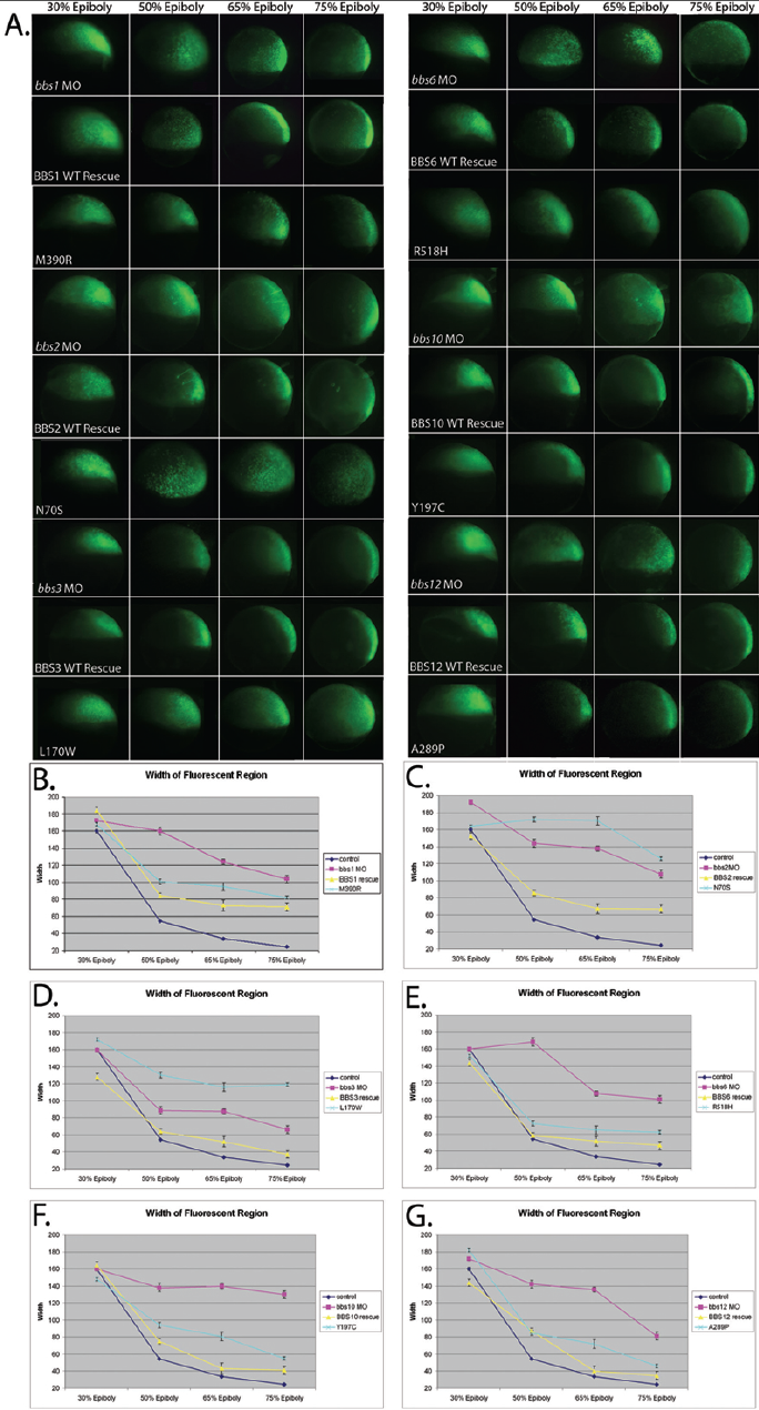
Fig. S3
Quantification of gastrulation defects. (A) Movement of cells during gastrulation stages is defective in MO-injected and null or dominant negative mutant rescued embryos, whereas WT rescued embryos are similar to uninjected controls. (B-G) The extent of gastrulation movements was quantified by measurement of the width of the field of fluorescing cells, repeated in triplicate – error bars shown. Dominant negative mutations, such as BBS2 N70S (C) and BBS3 L170W (D), produce cell movement defects more severe than MO alone, while null mutations such as BBS10 Y197C (F) are similar to MO. Hypomorphic mutations, such as BBS1 M390R (B) and BBS12 A289P (G) are less severe than MO, but do not efficiently rescue the gastrulation movement phenotype.