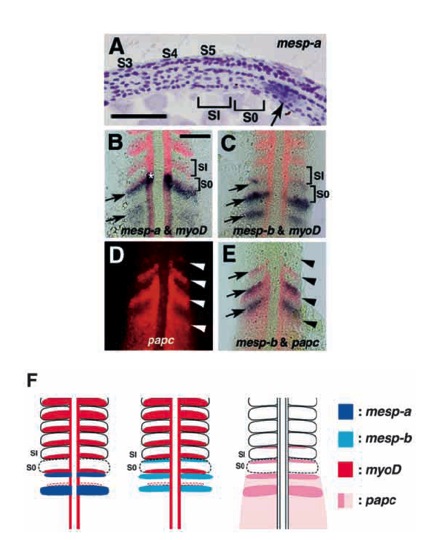
Fig. 3 Expression patterns of mesp-a and mesp-b in the presomitic mesoderm. Embryos are oriented with anterior to the top (except for A, in which anterior is to the left). (A) Longitudinal section through a 5-somite stage embryo hybridized with a mesp-a probe. Somites 3, 4 and 5 are labeled as S3, S4 and S5. A newly formed and forming (or the most anterior presumptive) somites are designated as SI and S0, respectively. The mesp-a-positive region (arrow) is located posterior to S0. (B,C) Two-colour staining with myoD (red) and mesp-a (blue in B) or mesp-b (blue in C) at the 10-somite stage. Dorsal views of flat-mounted embryos are shown. Arrows indicate the stripes of mesp genes. (D,E) Two-colour staining with paraxial protocadherin (papc, red) and mesp-b (blue) at the 10-somite stage. Dorsal views under fluorescence (D) and bright-field optics (E) are shown. Arrowheads indicate the anterior borders of papc expression domains and arrows indicate mesp-b expression stripes. Note that both expression domains overlap, sharing the same anterior border. (F) Simplified diagrams illustrating expression patterns of mesp-a, mesp-b, myoD and papc. Bars, 50 μm.
Image
Figure Caption
Figure Data
Acknowledgments
This image is the copyrighted work of the attributed author or publisher, and
ZFIN has permission only to display this image to its users.
Additional permissions should be obtained from the applicable author or publisher of the image.
Full text @ Development

