
|
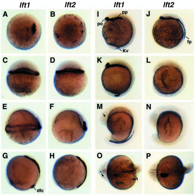
Fig. 2 lefty expression during early zebrafish development. (A-P) In situ localization of lft1 and lft2 transcripts at blastula through late somite stages (A,B, animal pole views; C-N, lateral views, dorsal at right; O,P, dorsoanterior views, anterior at left). lft1 and lft2 localize to the blastoderm margin at sphere (A,B) and dome stages (C,D). At shield stage (E,F), and at 90% epiboly (G,H) both genes localize to the dorsal hypoblast; lft1 is also expressed in dorsal forerunner cells (dfc). 1- to 3-somite embryos express both genes in the polster (po) and prechordal plate (pp, I,J); lft1 is uniquely expressed in Kupffer’s vesicle (Kv) (I), lft2 is uniquely expressed in floorplate precursors (fp) (J). At 6-8 somites lft1 expression is maintained (K) while lft2 is downregulated (L). 13-15 somite embryos express lft1 in the posterior notochord and left habenula (arrowhead) (M); lft2 is not expressed (N). 22-24 somite embryos showing lft1 in the anterior notochord (an), left habenula (arrowhead) and left lateral plate mesoderm (lpm) (O); lft2 is expressed in left lateral plate mesoderm (P).