
|
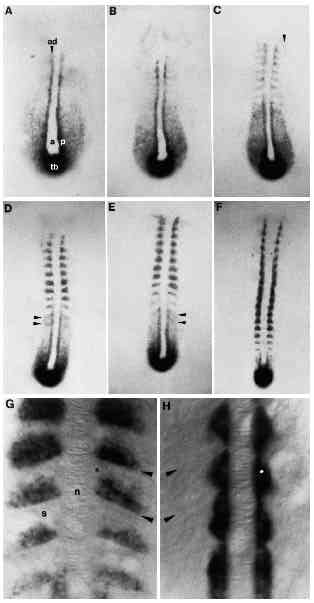
Fig. 4 Change in the snail1 expression pattern during somitogenesis. (A) 3-somite stage. snail1 RNA is intensively stained in adaxial cells (ad), a single row of paraxial hypoblast cells (p) adjacent to axial mesoderm (a). Lateral cells are also labelled but in a diffuse manner. The tail bud (tb) is strongly stained. (B) 4-somite stage. Labelling adjacent to the notochord starts to fade posterior to the furrow separating the third and fourth somite. Staining increases just anterior to each newly formed somitic furrow. (C) 7-somite stage. In the segmented mesoderm, signal disappears from adaxial cells. Note the epithelial character of adaxial cells more posteriorly in the unsegmented mesoderm. snail1 RNA is detected anterior to the somitic furrow in a single row of cells. See arrow showing snail1 RNA transiently expressed in the most lateral part of the segmental paraxial hypoblast. (D,E) 10- and 12- somite stages, respectively. In the unsegmented paraxial mesoderm (the segmental plate), there are two stripes of snail1 RNA at segment periodicity posterior to the most recently formed somite (see arrows). As each somite matures, the territory of snail1 expression spreads anteriorly within the somite. (F) 17-somite stage. In the most recently formed somite, snail1 RNA accumulates in a unique line of cells just anterior to the newly formed furrow. (G) Details of snail1 expression pattern in the somite (s) at 17- somite stage, dorsal view. Note the position of the notochord (n) in the middle and the location of the somitic furrow (arrows). snail1 RNA is detected in the posterior compartment of the somite. In the most recently formed somite, a single sheet of cells accumulates snail1 transcript. In older somites, the labelling occupies more of the posterior portion of the somite. The star indicates the position of muscle pioneer precursors which are not labelled with snail1 RNA. (H) 17-somite stage. Dorsal view of an embryo probed with a-tropomyosin. The arrow indicates the position of the somitic furrow. atropomyosin is detected in adaxial cells and in particular, in muscle pioneer precursors (indicated by a star). Note morphogenetic changes in a-tropomyosin-expressing cells as somites mature.