
Fig. 1
- ID
- ZDB-IMAGE-140107-40
- Genes
- Publication
- Reichert et al., 2013 - A BMP regulatory network controls ectodermal cell fate decisions at the neural plate border
- All Figures
- Figures for Reichert et al., 2013
|
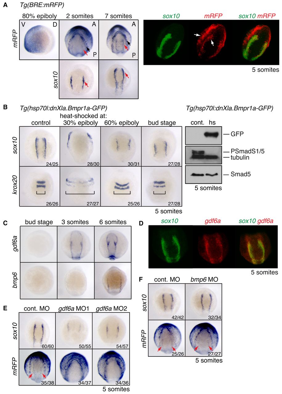
Fig. 1 Distinct domains of BMP activity exist at the NPB. (A) (Left) In situ hybridisation (ISH) for mRFP and sox10 in Tg(BRE:mRFP) zebrafish embryos. The 80% epiboly image is a lateral view, ventral to the left. Somitogenesis stages are shown in dorsal anterior view. Black arrow, outer domain of BMP activity; red arrow, inner NC domain. (Right) Double-fluorescent in situ hybridisation (DFISH) using probes against mRFP (Fast Blue, red) and sox10 (fluorescein-TSA, green). Arrows indicate the two distinct domains of BMP activity. Dorsal view, with anterior top right. (B) (Left) sox10 and krox20 ISH on Tg(hsp70l:dnXla.Bmpr1a-GFP) embryos heat shocked at the indicated stages. Brackets indicate the width of the neural ectoderm marked by krox20. (Right) Western blots of 5-somite stage Tg(hsp70l:dnXla.Bmpr1a-GFP) embryos heat shocked (hs) at bud stage, using the antibodies indicated. The control (cont.) corresponds to sibling embryos that were not heat shocked. (C) ISH for gdf6a and bmp6 at the stages indicated. (D) DFISH for sox10 and gdf6a. Dorsal view, with anterior to top. (E,F) Wild-type or Tg(BRE:mRFP) embryos were injected with 10 ng control MO or MOs against gdf6a (E) or bmp6 (F) and stained for sox10 or mRFP. Red arrows indicate the inner domain of BMP signalling as visualised by mRFP. The number of embryos with the observed phenotype among total embryos examined for a representative experiment is given bottom right. V, ventral; D, dorsal; A, anterior; P, posterior.