
Fig. 1
- ID
- ZDB-IMAGE-131218-46
- Genes
- Publication
- Cavodeassi et al., 2013 - Eph/Ephrin signalling maintains eye field segregation from adjacent neural plate territories during forebrain morphogenesis
- All Figures
- Figures for Cavodeassi et al., 2013
|
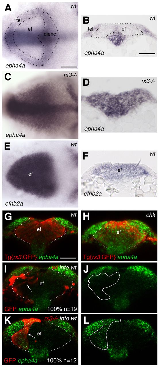
Fig. 1 Complementary expression of Ephrins and Ephs in the anterior neural plate (ANP) is lost in zebrafish rx3-/- mutants. (A-F) Whole-mount in situ hybridisations showing the expression in the ANP of epha4a in wild-type (A,B) and rx3-/- (C,D) embryos and of efnb2a in wild-type embryos (E,F). (G,H) Whole-mount in situ hybridisation to detect epha4a expression in the ANP (green) of Tg{rx3:GFP} (G) and Tg{rx3:GFP}; rx3-/- (chk) (H) embryos counterstained for GFP to highlight the eye field (red). (I-L) Transplants of wild-type (I,J) or rx3-/- (K,L) cells (labelled by GFP, red) into wild-type hosts. rx3-/- cells show autonomous activation of epha4a in the eye field (K,L). Arrows (I,K) point to transplanted cells. Dashed/dotted lines outline ANP domains (A), outline the eye field (B,G-I,K) or outline the transplants (J,L). All panels show frontal views with dorsal to the top of 1- to 3-somite stage (ss) embryos, except (A,C,E) which show dorsal views with anterior to the left. ef, eye field; tel, telencephalon; dienc, diencephalon. Scale bars: 50 μm.