
Fig. S4
- ID
- ZDB-IMAGE-131211-33
- Publication
- Phng et al., 2013 - Filopodia are dispensable for endothelial tip cell guidance
- All Figures
- Figures for Phng et al., 2013
|
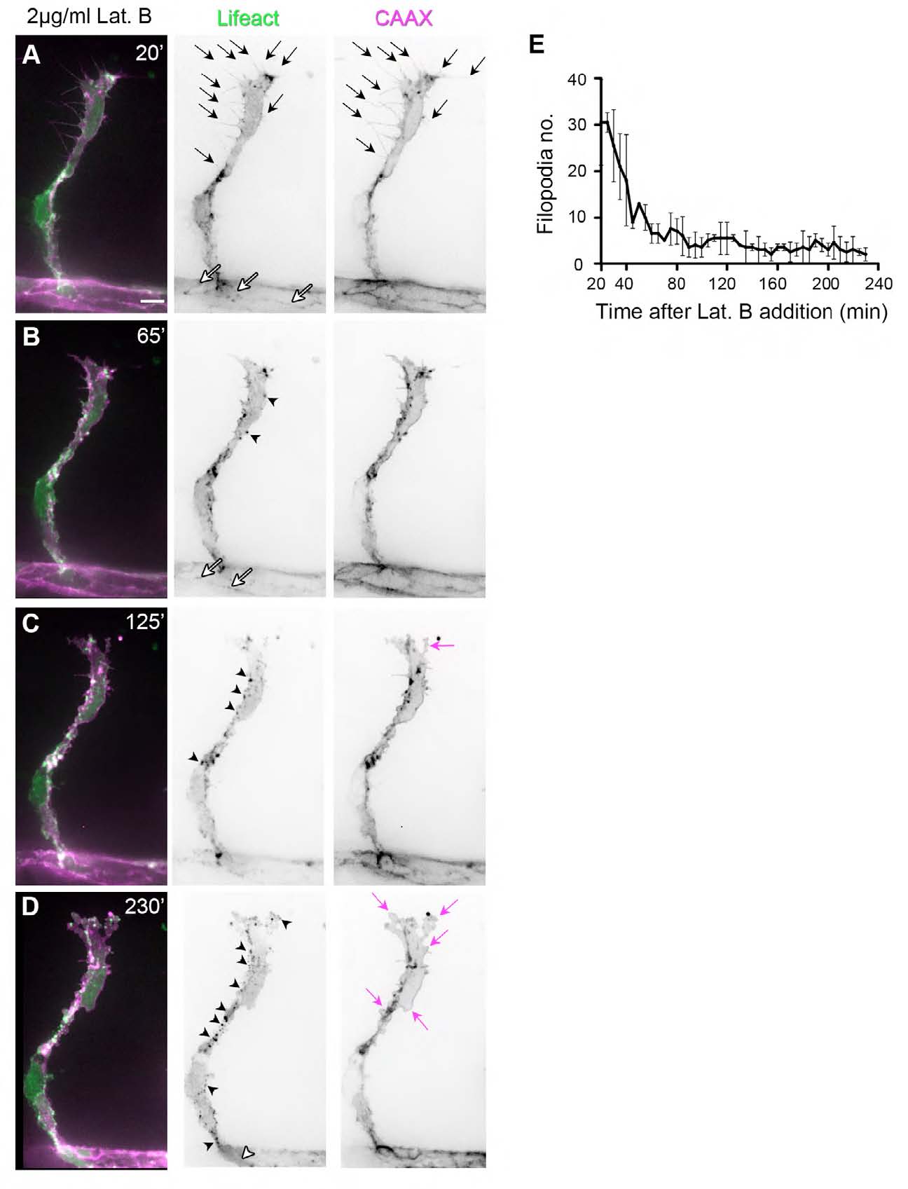
Fig. S4 Latrunculin B disrupts F-actin polymerisation and endothelial cell morphology. Tg(Fli1ep:Lifeact-EGFP); Tg(Kdr-l:ras- Cherry) s916 28.5 hpf embryos were treated with 2 μg/ml Lat. B. (A) At 20 minutes post-addition of Lat. B, endothelial tip cells of ISVs display multiple F-actin-filled filopodia (arrows) and junctional F-actin at the dorsal aorta (white arrows). (B-D) As drug treatment continued, fewer filopodia protrusions are observed. There is an increase in F-actin-positive puncta (black arrowheads) and loss of junctional F-actin (white arrowhead in D). Concomitant to perturbation of F-actin polymerisation, there is a change in EC morphology. The cell membrane becomes more convoluted at the cell body and at cell protrusions (magenta arrows). (E) Number of CAAX-positive filopodia after addition of 2 μg/ml Lat. B. n=2 ISVs from two embryos.