Fig. 1
Fig. 1
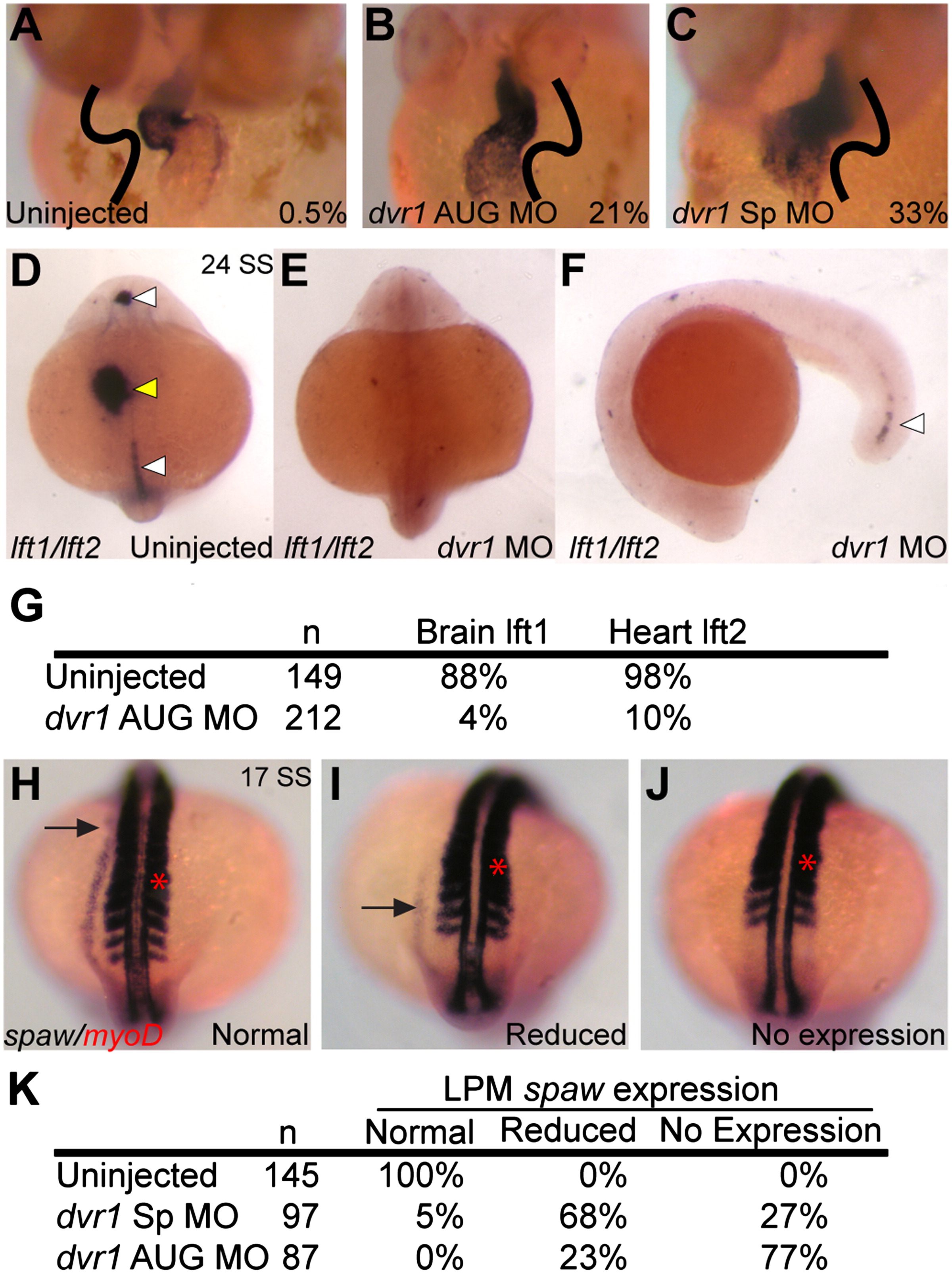
Knockdown of dvr1 in zebrafish causes heart, brain and LPM laterality defects. (A)–(C) Heart looping in control and morphant embryos, ventral view, cmlc2 in situ hybridization. Value in each panel indicates the percentage of reversed heart looping, orientation indicated by black lines. (A) Uninjected controls. (B) dvr1 AUG morphants. (C) dvr1 Sp morphants. (D)–(F) Asymmetric expression of lft1 in brain and expression in midline (white arrowheads) and asymmetric expression in lft2 in heart (yellow arrowhead) in control embryos (D) is lost in dvr1 morphants at 24 SS stage (E). Only a remnant lft1 expression is maintained in posterior midline (F). (G) Table showing quantification of brain lft1 and heart lft2 expression in both uninjected and dvr1 AUG morphant embryos. Values indicate percentage of normal expression. (H)–(J) Examples of different categories of spaw lateral plate mesoderm expression (black arrow) at 17 SS stage. myoD expression is also shown as a reference for spaw migration (red arrow). (H) Normal left-sided expression pattern of spaw. (I) Reduced spaw expression. (J) No spaw expression. (K) Quantification of spaw expression. In contrast to left-sided expression in control embryos, morphant embryos had reduced or no spaw expression in LPM.
Reprinted from Developmental Biology, 382(1), Peterson, A.G., Wang, X., and Yost, H. J., Dvr1 transfers left-right asymmetric signals from Kupffer's vesicle to lateral plate mesoderm in zebrafish, 198-208, Copyright (2013) with permission from Elsevier. Full text @ Dev. Biol.

