
|
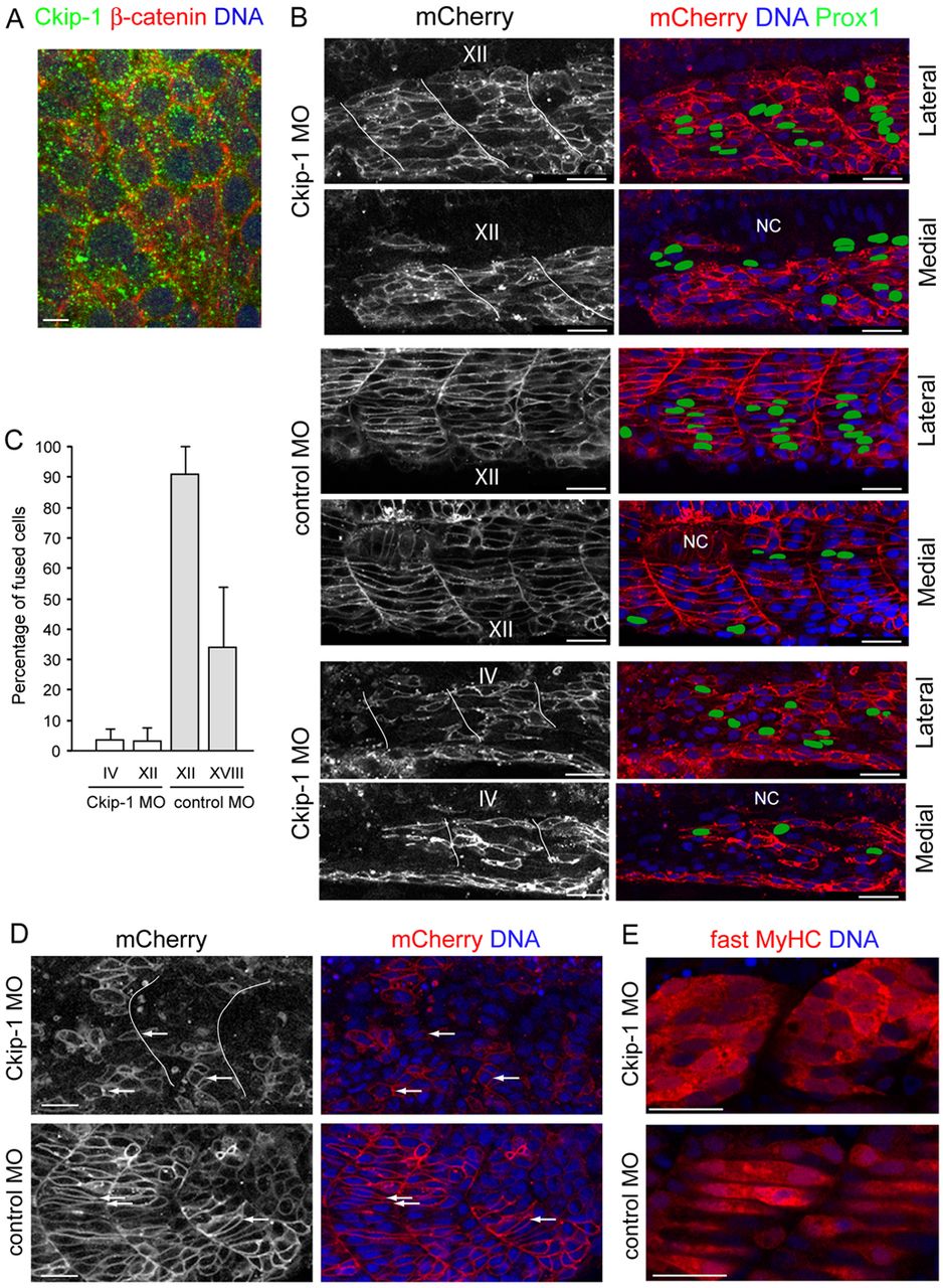
Fig. 2 Ckip-1 knock down impairs fast-twitch muscle precursor fusion. (A) Confocal image of Ckip-1 distribution in fast-twitch precursor cells, in an 8- to 10-somite embryo. Membranes were labelled using beta-catenin staining and nuclei with DAPI. Ckip-1 accumulates at the periphery of fast precursor cells. Dorsal view, anterior to the top. Scale bar: 5µm. (B) All panels (side view, anterior to the left) are confocal images showing myocytes in three somites, in 24 hpf embryos injected at the 1-cell stage with ckip-1 or control morpholinos and subsequently reinjected with membrane-mCherry mRNA at the 16- to 32-cell stage for membrane scatter labelling. Embryos were labelled with anti-Prox1 that stains slow fibre nuclei (green) and DAPI (blue). Prox1-positive nuclei were delineated and coloured green to be more visible. The Roman numerals above a somite indicate its positioning along the anterior–posterior axis (with I designating the anterior-most somite). Two images are shown for each embryo, one at the periphery of the myotome (lateral) and one at the level of the notochord (NC; medial). In morphant embryos, myosepta are delineated in white. Scale bars: 25µm. The membrane-mCherry staining was used to outline cells in confocal z-stacks (10–15 focal planes scanning the entire myotome), in order to count their nuclei. (C) Quantification of B, presented as the percentage of fused cells, i.e. containing more than one nucleus. Roman numerals under the bars indicate the analysed somite region. Values are means ± s.e.m. of six different morphant and control embryos. An average of 500 mCherry-positive cells was counted for each embryo. Whereas slow muscle fibres have reached the lateral myotome in the analysed regions (B), the fusion percentage is very low in ckip-1 morphants compared to controls, including in more anterior (more differentiated) somites (C). (D) Confocal images (mid-trunk, anterior to the left) encompassing a two-somite width view of 18 hpf embryos injected with Ckip-1-MO or control MO and mCherry mRNA, labelled with DAPI (blue). Arrows show short and elongating cells in 18 hpf Ckip-1 MO- and control-MO-injected embryos, respectively. Whereas in wild-type embryos, short fast muscle precursor cells elongate and intercalate along the anteroposterior axis, they remained irregular and most of them did not extend protrusions in the direction of elongation and between other cells. Scale bars: 25µm. (E) Confocal images showing a lateral view of two somites in 18 hpf Ckip-1-MO- and control-MO-injected embryos labelled for fast MyHC (F310; red) and DAPI (blue). Mid-trunk, anterior to the left. Scale bars: 25µm. Ckip-1 knock down does not interfere with differentiation.