
Image
|
Figure Caption
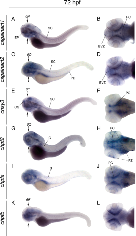
Fig. 5
Whole mount in situ hybridization of CS/DS glycosyltransferases at 72 hpf. Lateral views of larvae (left) and dorsal views of larval heads (right), showing the expression patterns of csgalnact1 (A,B), csgalnact2 (C,D), chsy3 (E,F), chpf2 (G,H), chpfa (I,J), and chpfb (K,L). Arrows indicate the position for sections shown in Figure 6. BVZ, brain ventrical zone; PC, pharyngeal cartilage; EP, ethmoid plate; G, gut; OS, olfactory system; PD, pronephric duct; PZ, proliferating zone of retina; SC, spinal cord.
Figure Data
Acknowledgments
This image is the copyrighted work of the attributed author or publisher, and
ZFIN has permission only to display this image to its users.
Additional permissions should be obtained from the applicable author or publisher of the image.
Full text @ Dev. Dyn.