
|
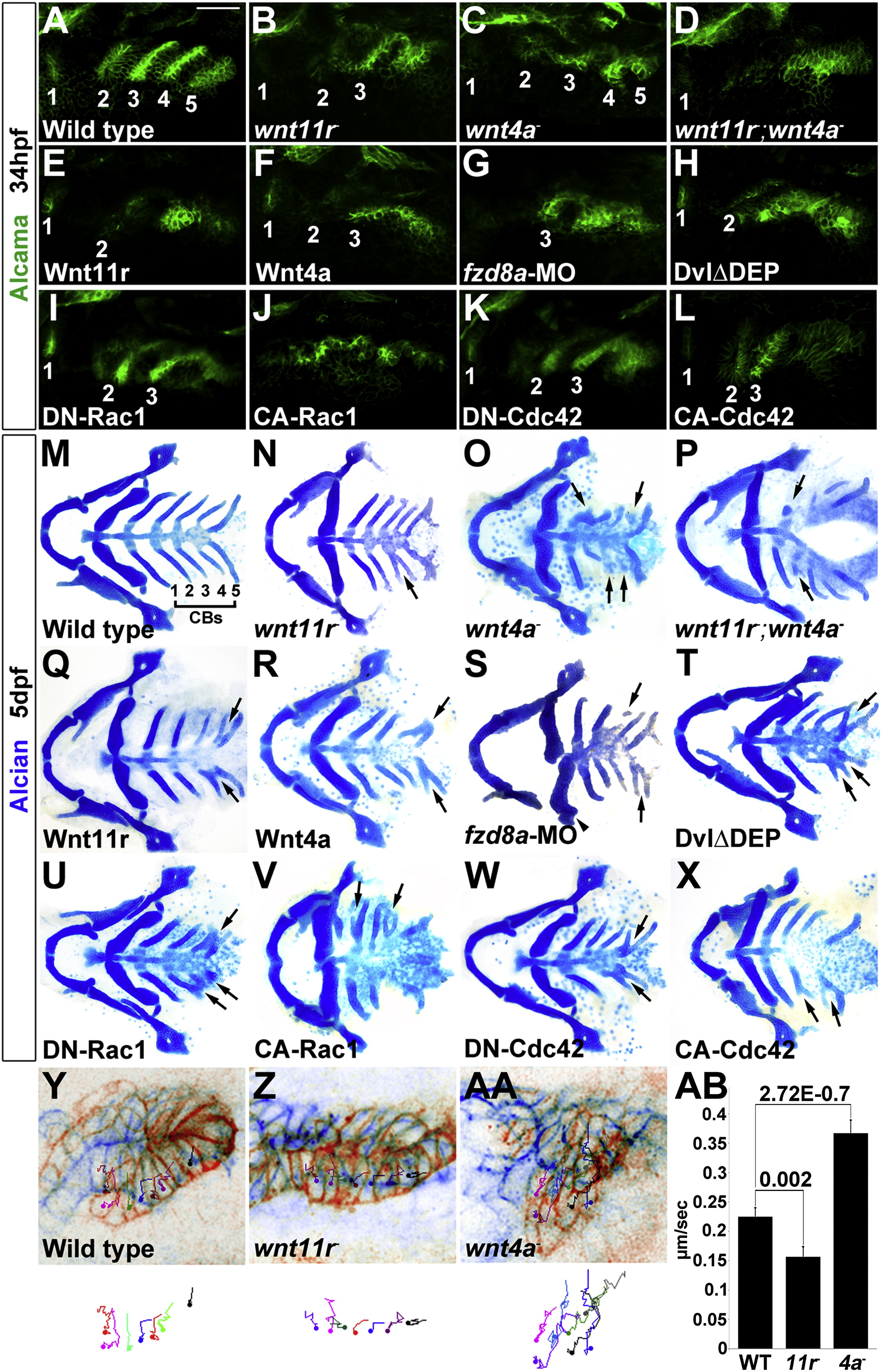
Fig. 3 Roles of Wnt Signaling Components in Pouch and CB Cartilage Development(A–L) Alcama immunohistochemistry (green) shows defects in pouches in mutant, fzd8a-MO, and transgenic embryos. Identifiable pouches are numbered. UAS transgenic embryos (capitalized) were doubly positive for nkx2.3:Gal4VP16. Scale bar, 40 μM.(M–X) Whole-mount views of dissected facial cartilages. Arrows indicated fused or abnormal CB cartilages. Arrowhead indicates abnormal hyoid cartilage in fzd8a-MO embryos.(Y–AA) Superimposition of initial (blue) and final (red) still images from time-lapse recordings of fifth pouch development in wild-type, wnt11r-/-, and wnt4a-/- embryos. Colored lines indicate cell tracks (shown also below merged images), with filled circles denoting final positions.(AB) Average pouch cell speed in wild-types and mutants. Data represent mean ± SEM and p values are shown for each comparison.See also Figures S1 and S2 and Movies S3, S4, and S5.
Reprinted from Developmental Cell, 24(3), Choe, C.P., Collazo, A., Trinh, L.A., Pan, L., Moens, C.B., and Crump, J.G., Wnt-Dependent Epithelial Transitions Drive Pharyngeal Pouch Formation, 296-309, Copyright (2013) with permission from Elsevier. Full text @ Dev. Cell