
Fig. 4
- ID
- ZDB-IMAGE-130117-19
- Publication
- Liu et al., 2012 - Prdm14 acts upstream of islet2 transcription to regulate axon growth of primary motoneurons in zebrafish
- All Figures
- Figures for Liu et al., 2012
|
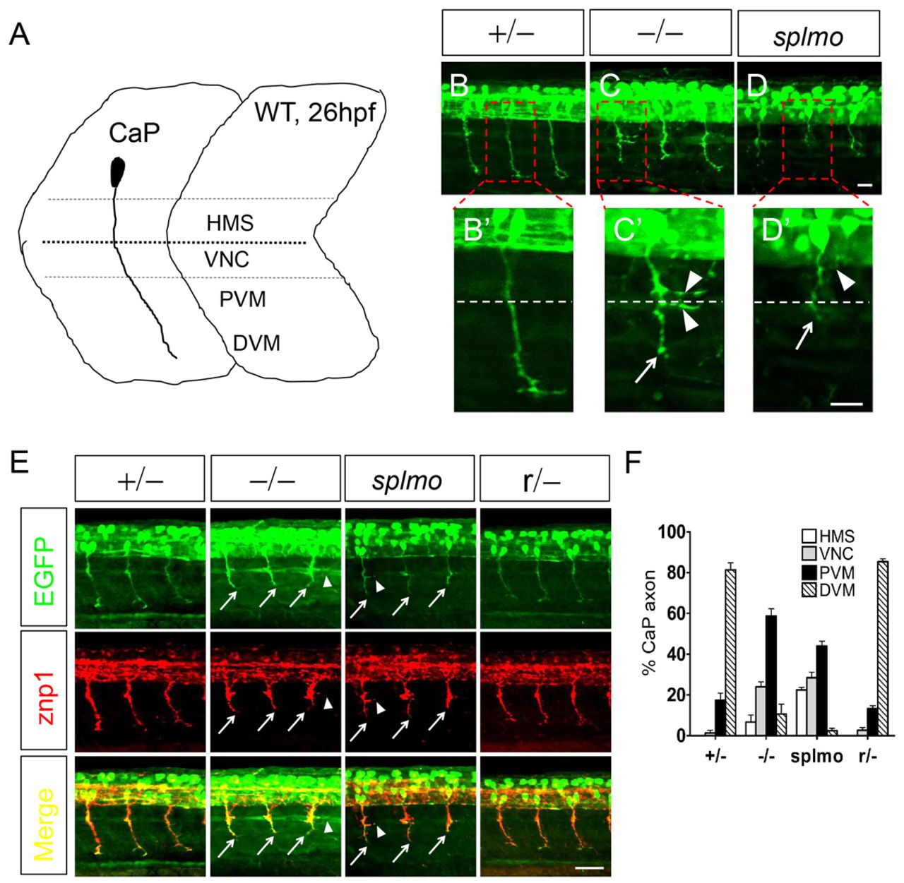
Fig. 4 CaP axons are shortened in slg mutant and splmo morphant zebrafish embryos. (A) CaP axon projection in 26-hpf wild type. Four regions are defined to describe CaP axon outgrowth: HMS, horizontal myoseptum; VNC, myotome adjacent to the ventral edge of the notochord; PVM and DVM, proximal and distal portion of the ventral myotome. (B-D2) slg mutant and splmo morphant embryos show greatly shortened CaP axons at 26 hpf. The boxed regions are magnified in B2-D2. Dashed line indicates horizontal septum. (E) znp1 immunostaining shows that the shortened axons are CaP in slg mutant and splmo morphant embryos. (F) Summary of CaP axon outgrowth (hemisegments 8-12) in slg heterozygous, slg homozygous, splmo morphant and slg revertant embryos. For each group, a total of 25 axons from five embryos were scored as HMS, VNC, PVM or DVM. Error bars indicate s.e.m. of triplicate experiments. Lateral views, dorsal to the top and anterior to the left. Arrows indicate shortened CaP axons; arrowheads indicate abnormal branched axons. Scale bars: 20 μm in B-D′; 50 μm in E.