Fig. 5
Fig. 5
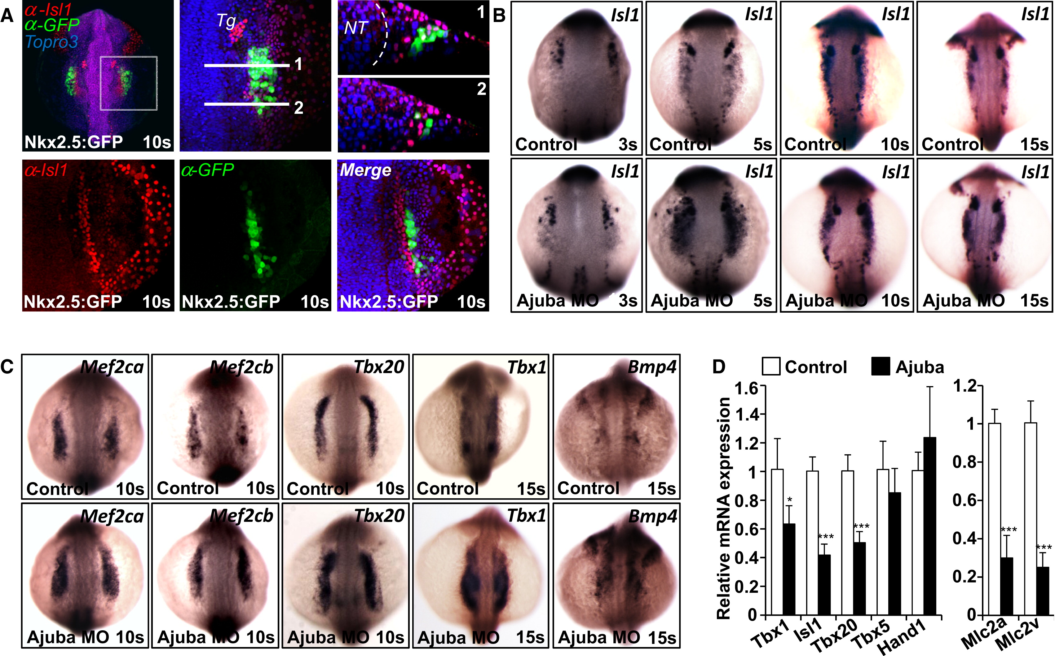
Ajuba Restricts the Number of SHF Progenitor Cells (A) Confocal images of Nkx2.5:GFP transgenic embryos stained with anti-Isl1 and anti-GFP antibodies at the 10 somite stage. The top-middle panel shows a higher magnification of the image in the top-left panel; the lines indicate the position of the transverse (xz) optical sections presented in top-right panel. NT, neural tube; Tg, trigeminal placode. (B) In situ hybridization of control and Ajuba morphant embryos at 3, 5, 10, and 15 somite stage with an Isl1 probe. (C) In situ hybridization of control and Ajuba morphant embryos with probes for Mef2ca, Mef2cb, Tbx20, Tbx1, and Bmp4. (D) Relative expression level of cardiac progenitor and cardiomyocyte marker mRNA normalized to GAPDH mRNA in day 5 EBs overexpressing either GFP only (control) or both GFP and Ajuba. Error bars represent SDs derived from two independent pools of control and Ajuba-overexpressing ES cells subjected to differentiation in EBs. See also Figures S4, S5, and S6.
Reprinted from Developmental Cell, 23(1), Witzel, H.R., Jungblut, B., Choe, C.P., Crump, J.G., Braun, T., and Dobreva, G., The LIM Protein Ajuba Restricts the Second Heart Field Progenitor Pool by Regulating Isl1 Activity, 58-70, Copyright (2012) with permission from Elsevier. Full text @ Dev. Cell

