
Fig. 9
- ID
- ZDB-IMAGE-120522-16
- Genes
- Publication
- Das et al., 2012 - Bmps and id2a act upstream of twist1 to restrict ectomesenchyme potential of the cranial neural crest
- All Figures
- Figures for Das et al., 2012
|
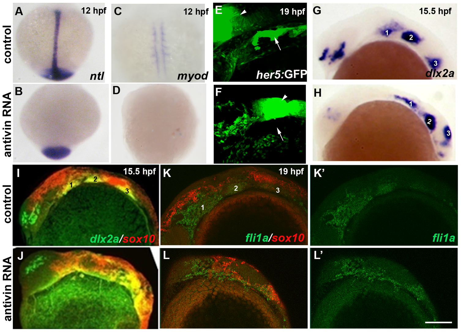
Fig. 9
Mesoderm and endoderm are not essential for ectomesenchyme formation.
(A–D) In situs at 12 hpf show loss of axial ntl and mesodermal myod expression in Antivin-mRNA-injected embryos compared to un-injected controls. (E,F) Confocal projections of her5:GFP expression at 19 hpf show loss of pouch endoderm (arrows) but not brain (arrowheads) in Antivin-mRNA-injected embryos compared to un-injected controls. (G,H) In situs at 15.5 hpf show normal ectomesenchyme induction of dlx2a in Antivin-mRNA-injected embryos (n = 12) and un-injected controls (n = 13). Arches are numbered. (I,J) Double fluorescent in situs at 15.5 hpf show normal induction of dlx2a (green) in sox10-positive CNCCs (red, yellow indicates co-localization) in Antivin-mRNA-injected embryos (n = 15) and un-injected controls (n = 9). (K,L) Double fluorescent in situs at 19 hpf show complementary expression of fli1a (green) in ectomesenchyme and sox10 (red) in non-ectomesenchyme in Antivin-mRNA-injected embryos (n = 5) and un-injected controls (n = 8). Scale bar = 50 μm.