
|
Fig. 8
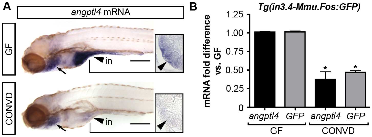
The intestinal module in3.4 recapitulates microbial suppression of angptl4.
(A) Semi-quantitative whole mount in situ hybridization of angptl4 mRNA in 6 dpf germ-free (GF) and conventionalized (CONVD) animals. Arrowheads mark intestinal expression. Note that the background staining in the gills (arrows) is similar in GF and CONVD fish. Transverse sections show that microbial suppression of angptl4 mRNA is specific to the intestinal epithelium. (B) Quantitative RT-PCR of angptl4 and GFP mRNA levels in 6 dpf GF and CONVD Tg(in3.4-Mmu.Fos:GFP) animals. GF and CONVD animals were derived from the same Tg(in3.4-Mmu.Fos:GFP) stable line. GFP and angptl4 mRNA were normalized to 18S rRNA levels and are shown as fold difference compared to GF controls averaged across 3 experimental replicates ± SEM (2 biological replicate groups of 10 larvae per condition per experiment). Similar results were attained when normalized to ribosomal protein L32 (rpl32) rRNA levels. Asterisks denote P-value<.01 from unpaired T-test between GF and CONVD conditions for each gene. See also Figure S8.