
Fig. 4
- ID
- ZDB-IMAGE-120405-59
- Genes
- Publication
- Miyake et al., 2012 - Neucrin, a novel secreted antagonist of canonical Wnt signaling, plays roles in developing neural tissues in zebrafish
- All Figures
- Figures for Miyake et al., 2012
|
Fig. 4
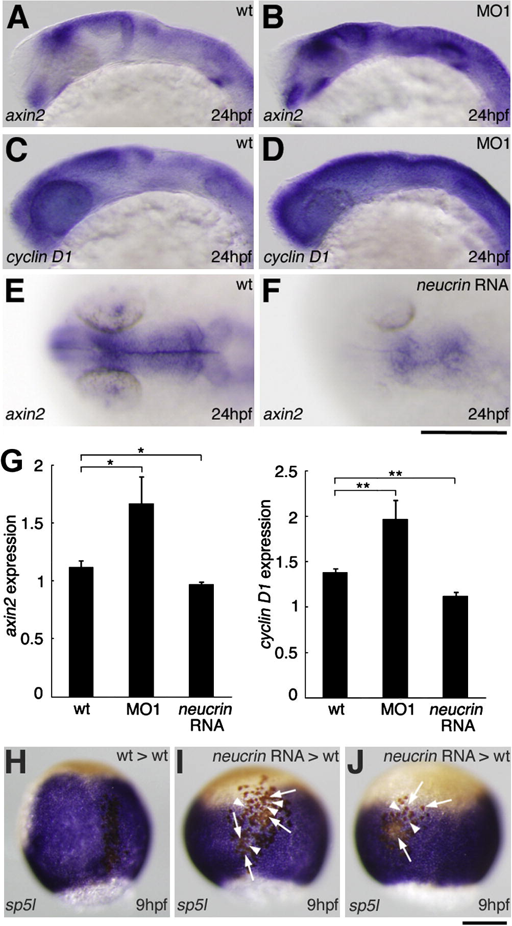
Effect of neucrin on the expression of Wnt/β-catenin target genes. (A-D) The expression of axin2 (A, B) and cyclin D1 (C, D) in wild-type (A, C) and neucrin MO1-injected (B, D) embryos at 24 hpf. Lateral view with anterior to the left. (E, F) The expression of axin2 in wild-type (E) and neucrin RNA-injected (F) embryos at 24 hpf. Dorsal view with anterior to the left. (G) qRT-PCR analysis on mRNA generated from wild-type embryos and embryos injected with neucrin MO1-injected and neucrin RNA-injected embryos. Results are the means ± S.D. (n = 3). The statistical significance of differences in mean values was assessed with the Student’s t-test. Asterisks indicate statistical significance compared with the wild type (*P < 0.05; **P < 0.01). (H-J) Dorsal view of sp5l expression in a wild-type embryo in which wild-type cells (H) or neucrin-overexpressing cells (I, J) were transplanted. Transplanted cells are labeled in brown. neucrin-overexpressing donor cells transplanted into the axis (I) and non-axial mesoderm (J) did not express sp5l (white arrows) and suppressed sp5l expression in the surrounding host regions (white arrowheads) at 9 hpf. Scale bar: 100 μm.
Reprinted from Mechanisms of Development, 128(11-12), Miyake, A., Nihno, S., Murakoshi, Y., Satsuka, A., Nakayama, Y., and Itoh, N., Neucrin, a novel secreted antagonist of canonical Wnt signaling, plays roles in developing neural tissues in zebrafish, 577-590, Copyright (2012) with permission from Elsevier. Full text @ Mech. Dev.