
Fig. 4
- ID
- ZDB-IMAGE-111223-24
- Genes
- Antibodies
- Publication
- Herwig et al., 2011 - Distinct Cellular Mechanisms of Blood Vessel Fusion in the Zebrafish Embryo
- All Figures
- Figures for Herwig et al., 2011
|
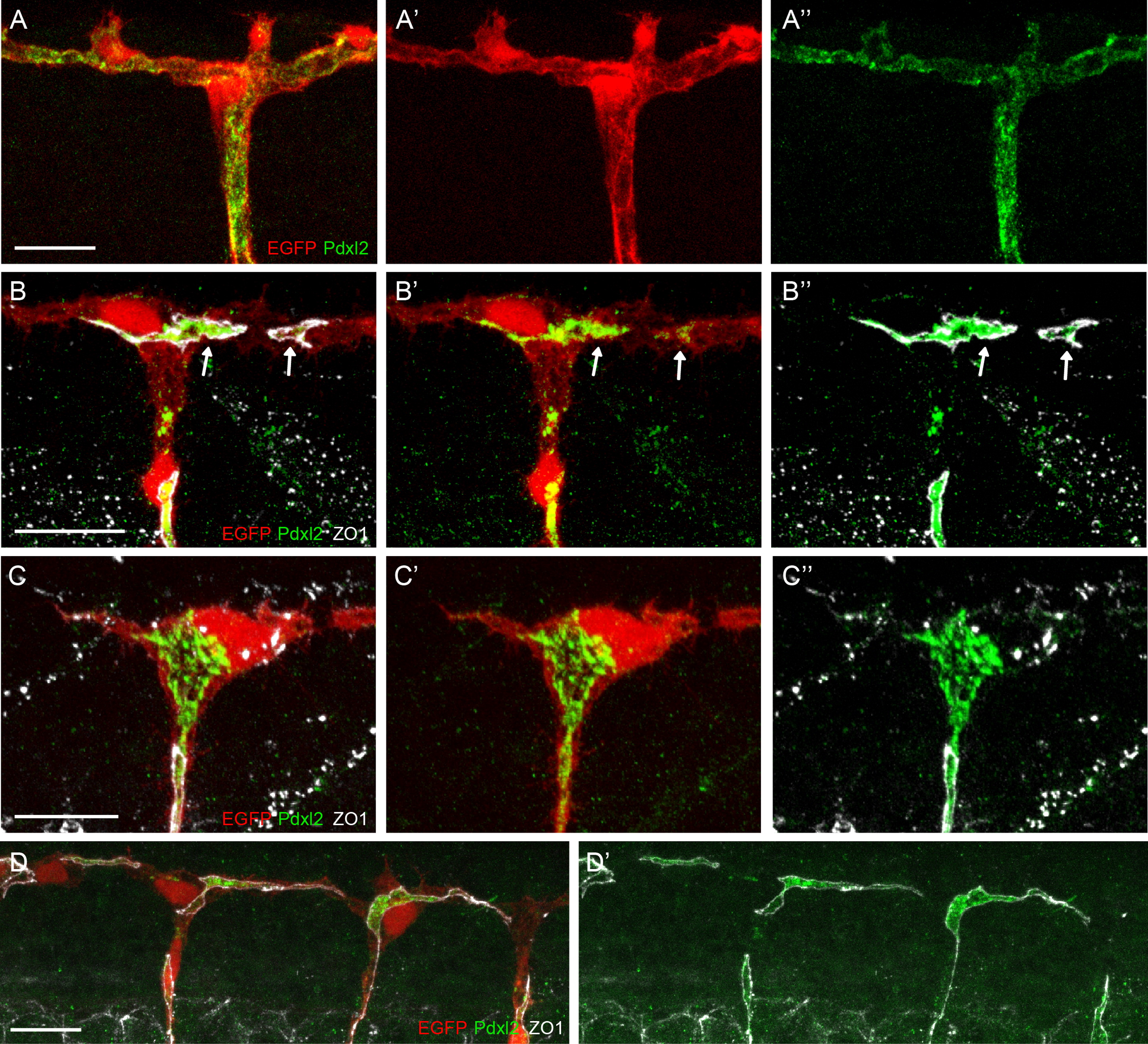
Fig. 4 Visualization of Apical Membrane during Anastomosis
(A–A′′) Pdxl2 antibodies label apical cell membranes in ISVs and DLAV at 48 hpf.
(B–B′′) Pdxl2 is localized within a junctional ring (arrow) at the contact site of two tip cells at the onset of anastomosis (36 hpf).
(C–C′′) Apical membrane invaginating into a tip cell at 36 hpf is shown by Pdxl2 immunostaining.
(D–D′) In sih morphants at the same stage, the Pdxl2 staining is only seen within the junctional rings, indicating that apical membrane invagination does not take place in the absence of blood flow.
Also compare Movie S3 and Figure S3A. α-ZO1 is shown in white, α-Pdxl2 in green, and kdrl:EGFP in red. Scale bar in all pictures represents 20 μm.