
IMAGE
Fig. S2
- ID
- ZDB-IMAGE-111215-13
- Genes
- Publication
- Oehlers et al., 2011 - The inflammatory bowel disease (IBD) susceptibility genes NOD1 and NOD2 have conserved anti-bacterial roles in zebrafish
- All Figures
- Figures for Oehlers et al., 2011
Image
|
Figure Caption
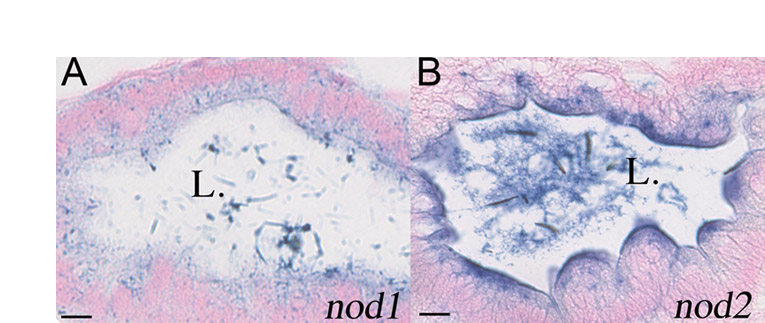
Fig. S2 Larval expression patterns of zebrafish nod1 and nod2. In situ hybridisation specimens of nod1 (A) and nod2 (B) sectioned and counterstained with Nuclear Fast Red. Photomicrographs are representative of intestinal bulb staining seen with antisense strand but not sense strand probes. Scale bars represent 10 μm, L. indicates lumen of the intestine.
Figure Data
Acknowledgments
This image is the copyrighted work of the attributed author or publisher, and
ZFIN has permission only to display this image to its users.
Additional permissions should be obtained from the applicable author or publisher of the image.
Full text @ Dis. Model. Mech.