Fig. 1
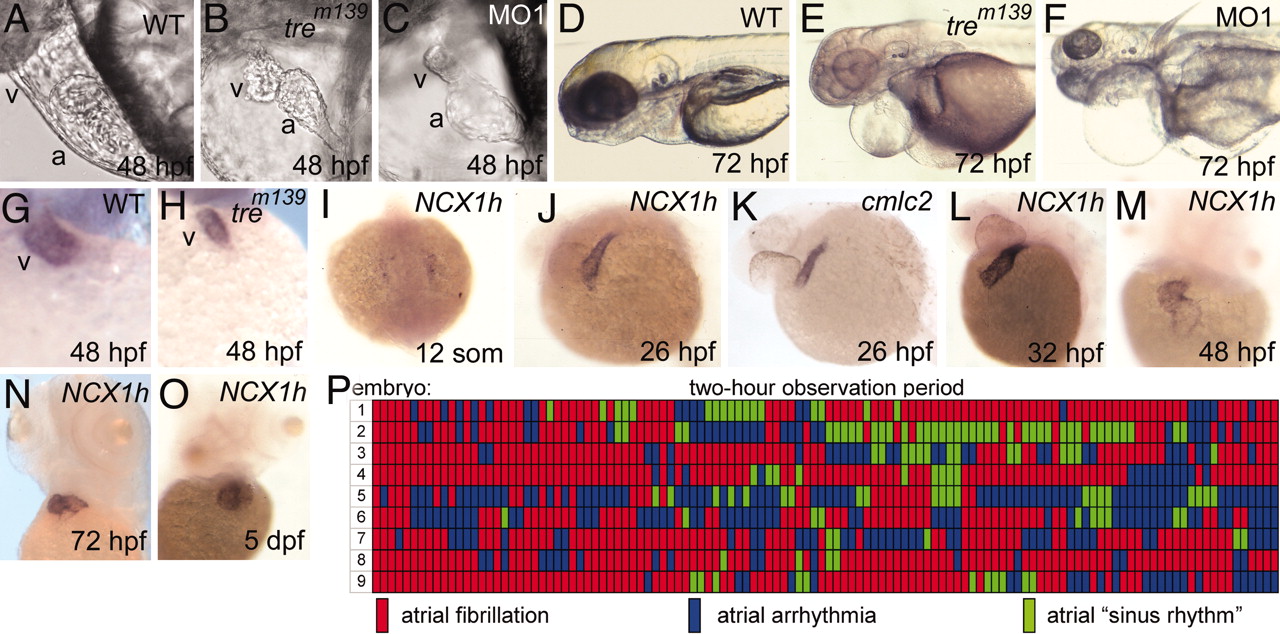
Cardiac defects in homozygous tre embryos. (A and B) Cardiac development at 48 hpf. The ventricle of tre mutants (B) is small and collapsed. (C) Injection of NCX1h-morpholino (MO1) phenocopies tre.(D–F) At 72 hpf, tre mutants and morphants show pericardial edema and slightly smaller heads. Heterozygous tre embryos (not shown) have no overt phenotype. (G and H) By 48 hpf, the ventricle, denoted by ventricle-specific vmhc expression, is markedly smaller in tre mutants. (I–O) NCX1h expression by in situ hybridization. (I) NCX1h is first detected in bilateral cardiac precursors at the 12-somite stage. (J and K) At 26 hpf, NCX1h expression in the heart tube is similar to cmlc2. NCX1h expression remains restricted to the heart at (L) 32 hpf, (M) 48 hpf, (N) 72 hpf, and (O) 5 days postfertilization. A second zebrafish NCX1 gene (NCX1b) is not expressed in the heart at any stage but is expressed in brain [see Langenbacher et al. (32)]. (P) Rhythm in the atria of nine tre embryos at 48 hpf depicted graphically. Each segment represents a 1-min observation period in which the atrial rhythm was visually scored in a single mutant embryo. Wild-type sibling embryos remained in sinus rhythm throughout the entire testing period (data not shown). No reproducible patterns in rhythm are observed on day 2 (P) or 3 (not shown) or in two other experimental replicates. a, atrium; v, ventricle.

