Fig. 6
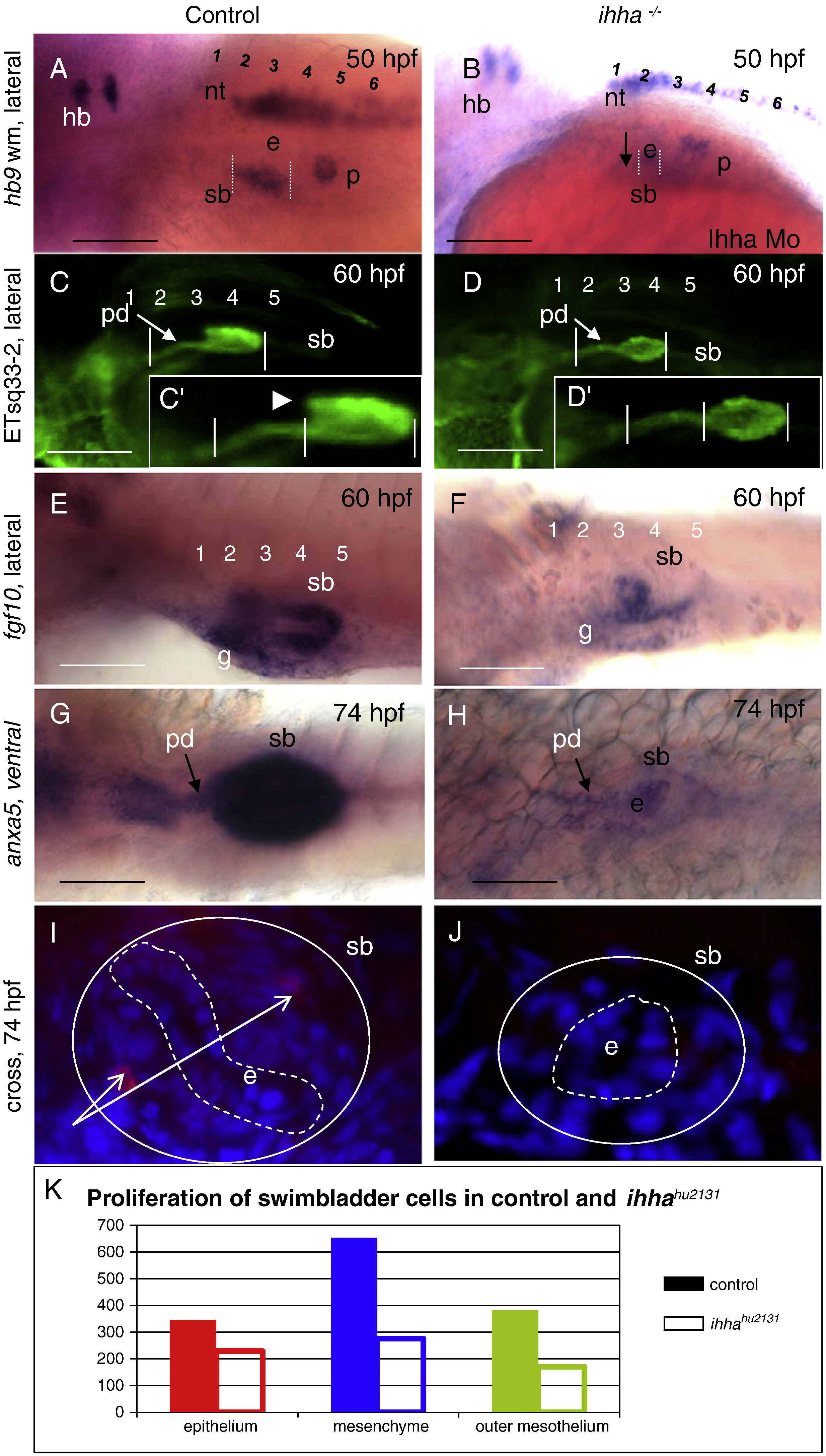
Defects of swimbladder development in the absence of Ihha. A, B, In control embryos expression of hb9, a marker for swimbladder epithelium, is initiated at 36 hpf and is expanded at 50 hpf. In Ihha morphant expression of hb9 in the swimbladder is delayed until 40–50 hpf, but expression of this marker elsewhere (the hindbrain, neural tube and endocrine pancreas) is unaffected. Note the same size of endocrine pancreas in control and Ihha morphant. C–D′, Swimbladder epithelia and pneumatic duct extensions (arrow) are significantly reduced in ihha-/-. Note that the anterior chamber (arrowhead) is absent in ihha-/- at 60 hpf. E, F, In control fgf10a-positive mesenchyme surrounded the epithelial bud starting from 48 hpf and increased at 60 hpf, in ihha-/- specification of swimbladder mesenchyme is initiated at around 60 hpf. G, H, Mesothelial domain of anxa5 in mutant larvae is almost absent, this staining also shows reduced swimbladder pneumatic duct and epithelium in mutant compared to control at 74 hpf. Anterior is to the left. I, J, Cross-section of swimbladder. HuC/D positive differentiated enteric neuron cell bodies are present in the mesenchymal layer of control larvae and absent along swimbladder mesenchyme in ihha-/-. Dotted white lines demarcate swimbladder epithelium. Solid white lines show the swimbladder size in control and ihha-/- larvae. K, assessment of cell numbers in swimbladder epithelium, mesenchyme and outer mesothelium of control and mutant. Solid colored bars correspond to control, while others correspond to ihha-/-. Abbreviations: hb, hindbrain; e, swimbladder epithelium; en, enteric neurons; p, endocrine pancreas; pd, pneumatic duct; n, notochord; nt, neural tube, s, somites; sb, swimbladder; g, gut. Numbers represent anterior somites.
Reprinted from Developmental Biology, 359(2), Korzh, S., Winata, C.L., Zheng, W., Yang, S., Yin, A., Ingham, P., Korzh, V., and Gong, Z., The interaction of epithelial Ihha and mesenchymal Fgf10 in zebrafish esophageal and swimbladder development, 262-76, Copyright (2011) with permission from Elsevier. Full text @ Dev. Biol.

