
Fig. 1
- ID
- ZDB-IMAGE-110915-1
- Genes
- Publication
- Zygmunt et al., 2011 - Semaphorin-PlexinD1 Signaling Limits Angiogenic Potential via the VEGF Decoy Receptor sFlt1
- All Figures
- Figures for Zygmunt et al., 2011
|
Fig. 1
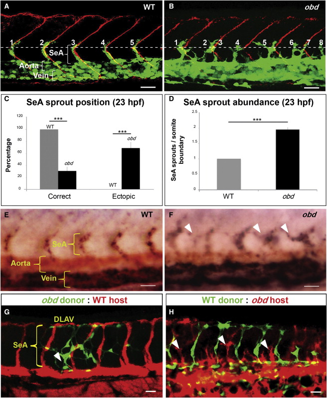
Sema-PlxnD1 Signaling Is Cell Autonomously Required within the Endothelium for Proper SeA Sprout Abundance and Distribution
(A and B) 23 hpf vasculatures, green. SBs, red. Horizontal myoseptum (HM), white dotted line. SeA sprouts, numbered. (A) WT. (B) obd.
(C and D) SeA sprout position (C) and abundance (D) in 23 hpf WT and obd. n = 8 WT, 12 obd. Error bars represent SEM. ***p < 0.001.
(E and F) WISH, 28 hpf trunks. Ectopic SeA sprouts, white arrowheads. Riboprobes: flt4 (blue), cdh5 (red). WT (E). obd (F). n = 10/10 WT, 10/10 obd.
(G and H) 32 hpf chimeric vasculatures with ECs of donor (green) and host (red) origin. Examples of ectopic SeA sprouts, white arrowheads.
(A, B, E–H) Anterior, left; dorsal, up. Scale bars represent 30 μm. See Figure S1.
Reprinted from Developmental Cell, 21(2), Zygmunt, T., Gay, C.M., Blondelle, J., Singh, M.K., Flaherty, K.M., Means, P.C., Herwig, L., Krudewig, A., Belting, H.G., Affolter, M., Epstein, J.A., and Torres-Vazquez, J., Semaphorin-PlexinD1 Signaling Limits Angiogenic Potential via the VEGF Decoy Receptor sFlt1, 301-314, Copyright (2011) with permission from Elsevier. Full text @ Dev. Cell