
Fig. 3
- ID
- ZDB-IMAGE-110812-25
- Genes
- Antibodies
- Publication
- Naganawa et al., 2011 - Developmental transition of touch response from slow muscle-mediated coilings to fast muscle-mediated burst swimming in zebrafish
- All Figures
- Figures for Naganawa et al., 2011
|
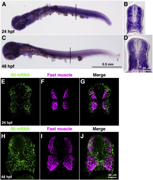
Fig. 3
flii is expressed in the CNS and muscle. (A–D) In situ hybridization with flii probe. flii appeared to be expressed ubiquitously in whole embryos at 24 hpf (A) and 48 hpf (C). Examination of cross sections confirmed that flii was expressed in muscle tissues (B,D). (E–J) Double labeling with flii mRNA and fast muscle myosin. Expression of flii was observed ubiquitously at both 24 hpf (E) and 48 hpf (H). Anti-fast myosin labeled fast muscle (F,I) but not the superficial slow muscle. In double labeling, flii expression was also seen outside the fast muscle (G,J), suggesting that flii was expressed in both slow and fast muscle.
Reprinted from Developmental Biology, 355(2), Naganawa, Y., and Hirata, H., Developmental transition of touch response from slow muscle-mediated coilings to fast muscle-mediated burst swimming in zebrafish, 194-204, Copyright (2011) with permission from Elsevier. Full text @ Dev. Biol.