
|
Fig. 2
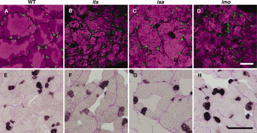
Characterization of premeiotic germ cells in the its, isa, and imo zebrafish mutants. A–D: Immunohistochemical analysis of Plzf. Fresh-frozen sections of wild-type (A), its (B), isa (C), and imo (D) testes were stained with anti-Plzf antibodies (green). The nuclei stained with TOPRO-3 are visible in a magenta pseudo-color. Plzf-expressing male zebrafish germ cell populations, which contain type A spermatogonia and early type B spermatogonia, were normal in terms of cell number and distribution patterns in each mutant. E–H: Analysis of cell proliferation by BrdU (5-bromo-22-deoxy-uridine) incorporation. Zebrafish were labeled with BrdU for 2 hr and testicular paraffin sections were subsequently stained with anti-BrdU antibodies and counterstained with periodic acid–Schiff. In the wild-type zebrafish testis, cysts of BrdU-positive type B spermatogonia, which are premeiotic spermatogenic cells, were observed. In the testis of the its (F), isa (G), and imo (H) mutants, the incorporation of BrdU in type B spermatogonia were also observed. Scale bars = 100 μm.