
Fig. 6
- ID
- ZDB-IMAGE-110622-91
- Genes
- Antibodies
- Publication
- Yang et al., 2011 - BMP and non-canonical Wnt signaling are required for inhibition of secondary tail formation in zebrafish
- All Figures
- Figures for Yang et al., 2011
|
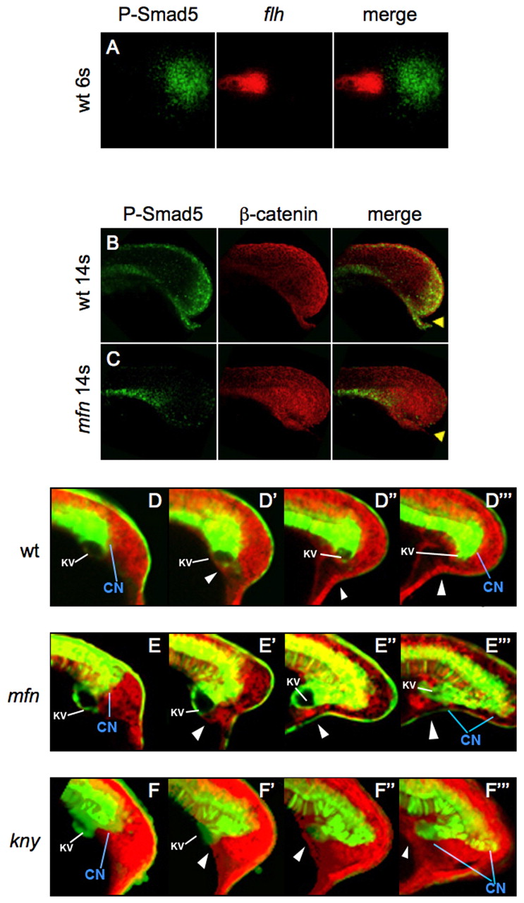
Fig. 6
Defects of the caudal end of notochord in mfn and kny initiate during tailbud protrusion. (A) Dorsal view of flat-mounted tailbud of a 6-somite wild-type zebrafish embryo, after fluorescence in situ hybridization (FISH) for flh (red) and antibody staining for P-Samd5 (green); no colocalization (merge) is seen. (B,C) Lateral view of tails of 14-somite wild-type and mfn embryos exposed to P-Smad5 (green) and β-catenin (red) antibodies. P-Smad5 staining is absent in the posterior tailbud of mfn (C, merge). Arrowheads indicate the ventroposterior cells in the tailbud that are normally positive for P-Smad5 (B, merge). (D-F4) Confocal time-lapse recording of tailbuds in embryos expressing Tg(flh:EGFP) transgene (green) and mRFP (red) from the 10-somite stage. Shown are wild type (D-D4) and mfn (E-E4) and kny (F-F4) mutants. Note that transgenic EGFP is retained in the caudal notochord, floorplate and periphery of Kupffer′s vesicle (KV; marked by the white line). The caudal end of notochord (CN) is indicated by blue line. Arrowheads mark the ventroposterior cells in the tailbud. Embryos were all mounted laterally.