
Fig. 4
- ID
- ZDB-IMAGE-110421-5
- Genes
- Publication
- Fujimoto et al., 2011 - Identification of a Dopaminergic Enhancer Indicates Complexity in Vertebrate Dopamine Neuron Phenotype Specification
- All Figures
- Figures for Fujimoto et al., 2011
|
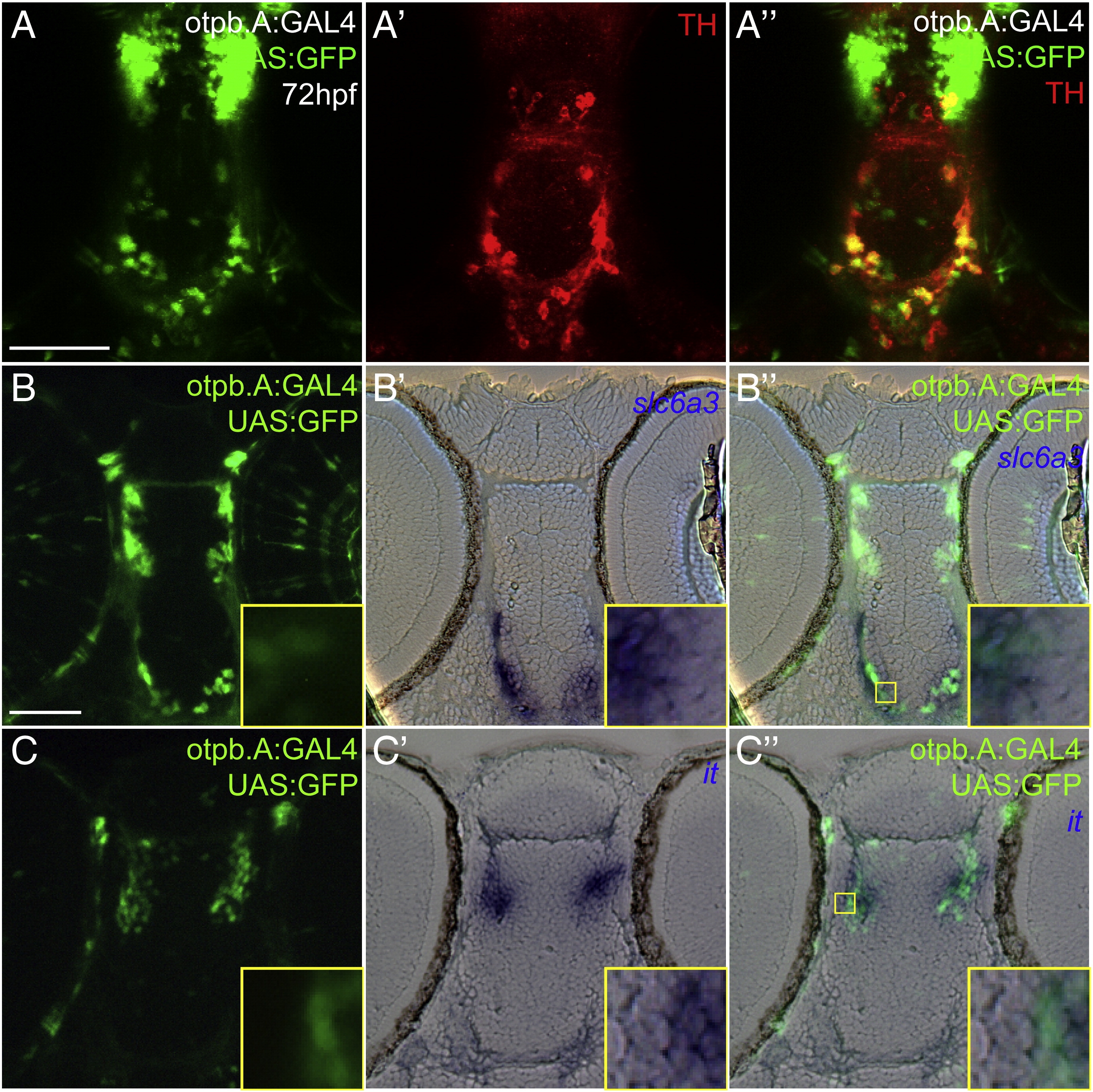
Fig. 4
Characterization of otpb.A expression in dopaminergic and neuroendocrine cells. Embryos at 72 hpf, Tg(otpb.A:GAL4)zc57, Tg(UAS:GFP). Scale bar is 50 μm. (A–A3) Confocal z-stack projection of whole-mount embryos double-labeled for GFP and for TH immunohistochemistry, ventral views, anterior to the top. (B) and (C) Plastic horizontal sections of double-labeled Tg(otpb.A:GAL4)zc57; Tg(UAS:GFP) embryos at 72 hpf, anterior to the top. Higher magnification inset showing co-localization is indicated by yellow boxed area and is shown at bottom right of each panel. (B–B3) Labeled for GFP antibody (B) and slc6a3 mRNA (B2), GFP-positive neurons in the diencephalon co-express slc6a3, confirming their identity as dopaminergic. (C–C3) Labeled for GFP antibody (C) and isotocin mRNA (C2), rostral diencephalon GFP-positive NPO neurons co-express GFP and isotocin.
Reprinted from Developmental Biology, 352(2), Fujimoto, E., Stevenson, T.J., Chien, C.B., and Bonkowsky, J.L., Identification of a Dopaminergic Enhancer Indicates Complexity in Vertebrate Dopamine Neuron Phenotype Specification, 393-404, Copyright (2011) with permission from Elsevier. Full text @ Dev. Biol.