
Fig. S5
- ID
- ZDB-IMAGE-110408-8
- Genes
- Publication
- Corti et al., 2011 - Interaction between alk1 and blood flow in the development of arteriovenous malformations
- All Figures
- Figures for Corti et al., 2011
|
Fig. S5
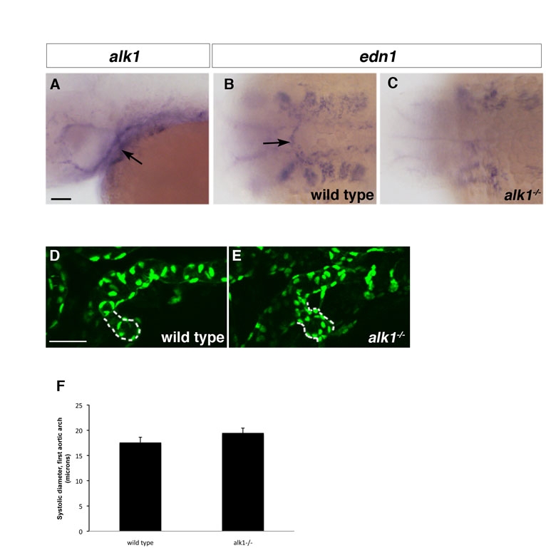
The first aortic arch is alk1 positive and abnormal in alk1 mutants. (A-C) In situ hybridization showing alk1 (A) and edn1 (B,C) expression in 36 hpf wild-type (A,B) and alk1-/- embryos. alk1 is expressed in the first aortic arch at 36 hpf (A, arrow). edn1 is expressed in the first aortic arch in wild-type embryos (B, arrow) but expression is lost in alk1 mutants (C). (D,E) Two-dimensional projections of confocal z-series 36 hpf Tg(fli1a:nEGFP)y7 embryos. The first aortic arch is enlarged in alk1 mutants (E) compared with wild-type siblings (D). However, at this early time, differences are not yet statistically significant (F). Data in F represent an average of three systolic diameters per embryo measured from high speed confocal micrographs, mean±s.e.m. n=8 wild type; n=5 alk1-/-. (A,D,E) Lateral views, anterior leftwards. (A,D) left side. (E) right side. (B,C) dorsal view, anterior leftwards. Scale bars: 50 μm.