
Fig. 1
- ID
- ZDB-IMAGE-110324-27
- Genes
- Antibodies
- Publication
- Sheets et al., 2011 - Ribeye is required for presynaptic CaV1.3a channel localization and afferent innervation of sensory hair cells
- All Figures
- Figures for Sheets et al., 2011
|
Fig. 1
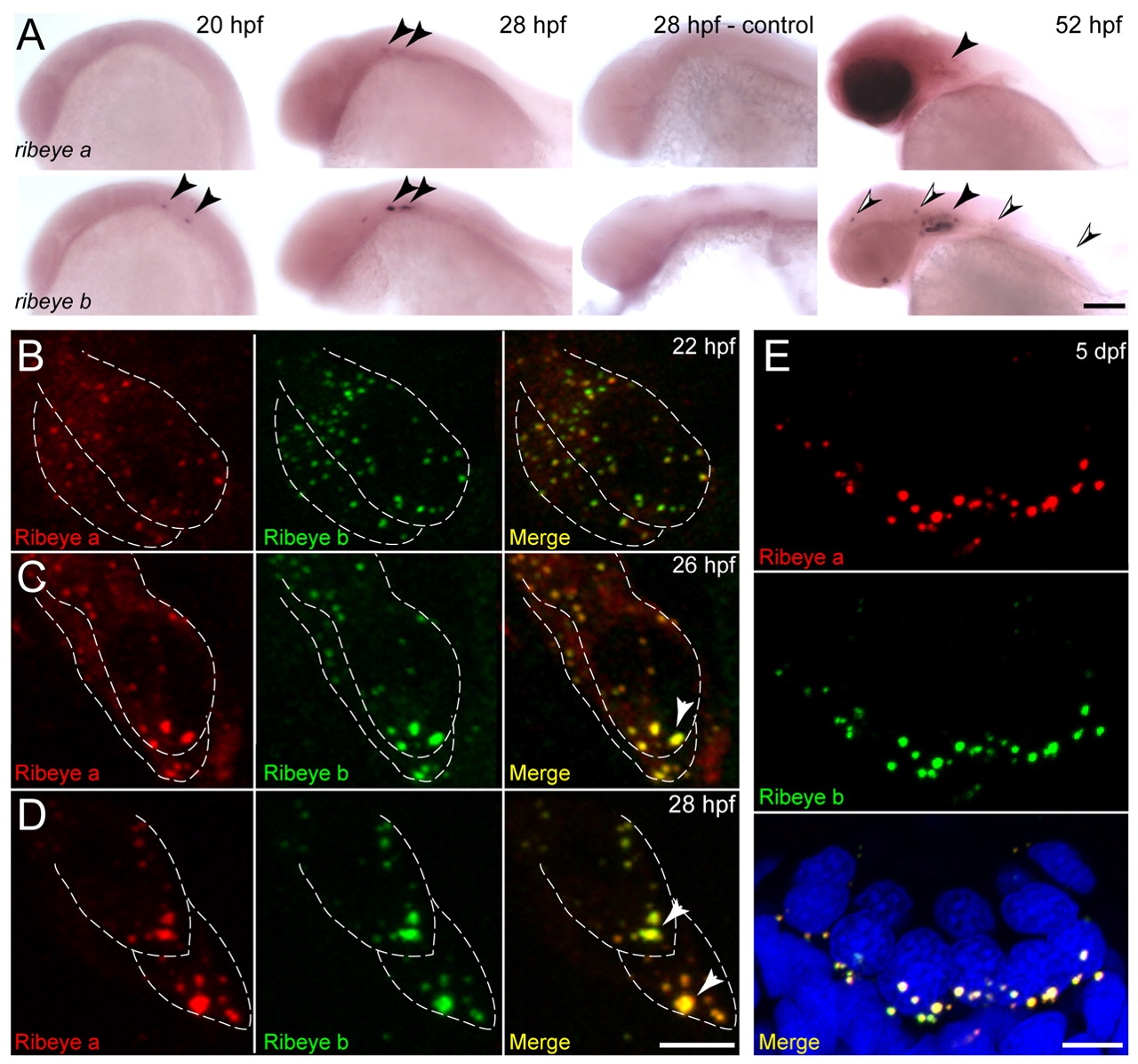
ribeye a and ribeye b expression and localization during zebrafish hair-cell development. (A) Expression of ribeye a and ribeye b mRNA in the developing ear and neuromasts. ribeye b is expressed strongly in the otic vesicle at 20 hpf, while ribeye a is expressed after 20 hpf, and at lower levels (black arrowheads). At 52 hpf, ribeye a is detectable in the retina and ear (black arrowhead), whereas ribeye b is expressed in both the ear (black arrowhead) and lateral line neuromasts (black and white arrowheads). Scale bar: 200 μm. (B-E) Representative maximum confocal z-projections of hair cells in wild-type larvae. Scale bars: 3 µm. (B-D) Ribeye immunofluorescent labeling in nascent hair cells in the developing ear at 22 hpf (B), 26 hpf (C) and 28 hpf (D). Ribeye a and Ribeye b proteins form aggregates that, by 26 hpf, are accumulated at the basal end of hair cells (white arrowheads). (E) Ribeye immunofluorescent labeling in a cross-section of a neuromast at 5 dpf. Merged images include DAPI labeling of cell nuclei (blue).