
IMAGE
Fig. 3
- ID
- ZDB-IMAGE-101206-6
- Genes
- Antibodies
- Publication
- Hilario et al., 2010 - Collagen XIXa1 is crucial for motor axon navigation at intermediate targets
- All Figures
- Figures for Hilario et al., 2010
Image
|
Figure Caption
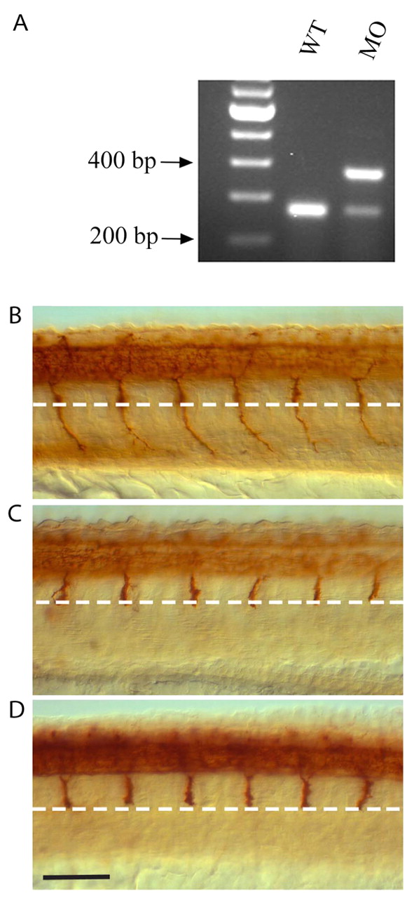
Fig. 3 ColXIX knock down phenocopies the stumpy mutation. (A) RT-PCR shows inclusion of a 109 bp fragment in splice-blocking colXIX MO injected (MO) compared to wild-type (wt) embryos. (B-D) CaP axon phenotypes as visualized using znp-1 antibody in (B) uninjected wild-type embryos, (C) wild-type embryos injected with 9 ng colXIX MO and (D) stumpyb393-/- mutant embryos. White dashed line indicates the horizontal myoseptum. Scale bar: 70 μm.
Figure Data
Acknowledgments
This image is the copyrighted work of the attributed author or publisher, and
ZFIN has permission only to display this image to its users.
Additional permissions should be obtained from the applicable author or publisher of the image.
Full text @ Development