
Fig. 1
- ID
- ZDB-IMAGE-101122-28
- Genes
- Publication
- Soza-Ried et al., 2010 - Essential role of c-myb in definitive hematopoiesis is evolutionarily conserved
- All Figures
- Figures for Soza-Ried et al., 2010
|
Fig. 1
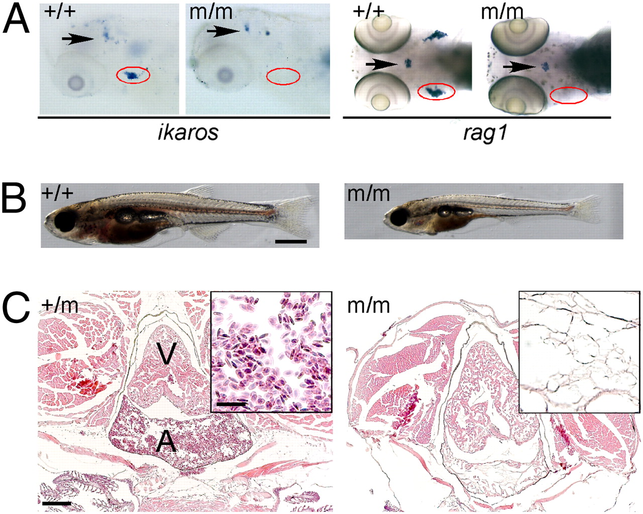
Phenotype of IP109 mutants. (A) Whole-mount RNA in situ hybridization of wild-type (+/+) and homozygous IP109 mutant (-/-) embryos with probes for ikaros (lateral views; Left) and rag1 (dorsal views; Right) at 5 d postfertilization (dpf). Note that ikaros is also expressed in neurons (arrows); this hybridization signal serves as an internal positive control for the hybridization process. In the hybridizations with rag1, a gh probe labels growth hormone-producing cells in the hypophysis (arrows) and serves as control for hybridization. The region of the thymus is encircled. No differences were seen between +/+ and +/m fish. Table S1 has probe details. (B) Macroscopic view of wild-type (Left) and mutant (Right) fish at 20 dpf. Note the smaller size and pale appearance of mutants. (Scale bar: 1 mm.) (C) Histological sections through the regions of the heart (A, atrium; V, ventricle) of heterozygous (Left) and homozygous mutant (Right) fish at 8 wk of age. Note the complete absence of erythrocytes in the mutant fish (Fig. S1). H&E staining. (Scale bars: 400 μm; Inset, 100 μm.)