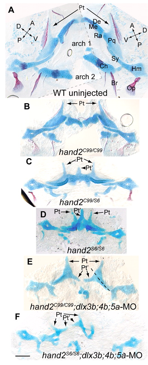
Fig. 7 hand2 mutants and hand2 mutants injected with dlx3b;4b;5a-MO show homeotic skeletal phenotypes. (A-F) Alcian Blue and Alizarin Red staining at 6 dpf. Images are flat mounted bilateral pharyngeal arches, oriented with midline to the center, and anterior upwards. (A) The wild-type skeleton was too large for a single image at this magnification, so two images were overlaid for this panel (border indicated with a broken grey line). (B) hand2C99 homozygotes have reduced ventral, but normal intermediate and dorsal domain skeleton. (C) In trans-heterozygous fish carrying hand2C99 and hand2S6, defects are typically more severe than in hand2C99 homozygotes, but less severe than in (D) hand2S6 homozygotes. In hand2S6 homozygotes, broad cartilages often span the midline, similar in shape to duplicated palatoquadrates, complete with pterygoid processes (arrows). (E) When hand2C99 homozygotes are injected with dlx3b;4b;5a-MO, joints are lost in both arches, and the remainder of Meckel′s cartilage is tapered out into a shape similar to a pterygoid process. A broken line indicates the first arch dorsal-ventral plane of symmetry. (F) The cartilage expansions of hand2S6 are lost when dlx3b;4b;5a-MO is injected. The palatoquadrate of hand2S6;dlx3b;4b;5a-MO is often severely defective, though the distance between the first and second arch-derived skeleton seen on the left side of F is exaggerated by mounting artifacts. Scale bar: 100 μm.
Image
Figure Caption
Figure Data
Acknowledgments
This image is the copyrighted work of the attributed author or publisher, and
ZFIN has permission only to display this image to its users.
Additional permissions should be obtained from the applicable author or publisher of the image.
Full text @ Development

