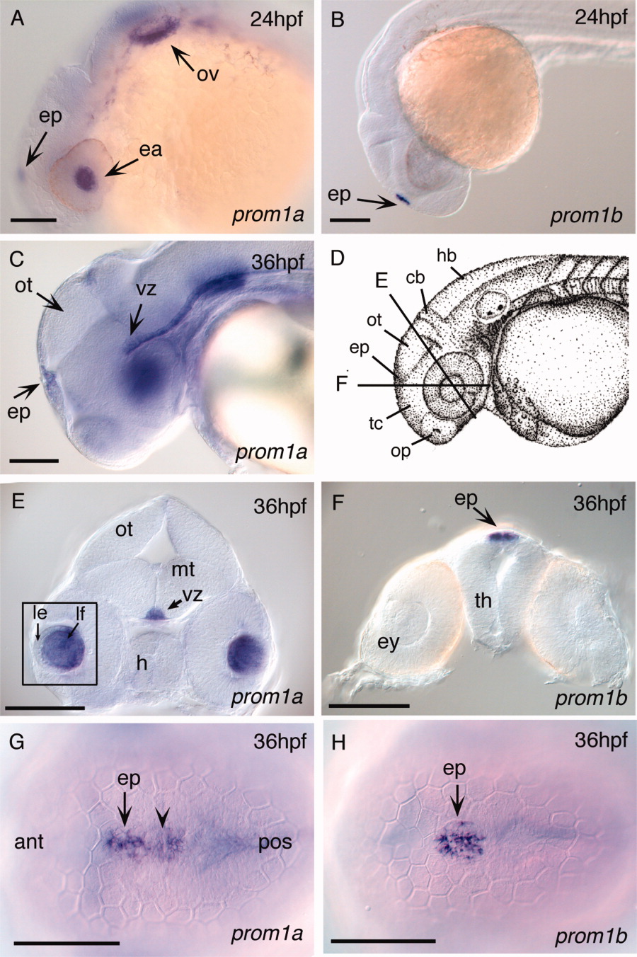
Fig. 3 Expression of zebrafish prom1a and prom1b in developing sensory organs and CNS of 24- and 36hpf embryos. A: Lateral view, 24hpf. prom1a expression in the lens anlage, epiphysis, and otic vesicle. B: Lateral view, 24hpf. prom1b expression in the epiphysis (arrowhead). C: Lateral view, 36hpf. prom1a expression in the ventricular zone along the dorsal surface of the midbrain tegmentum and extending into the hindbrain. D: Camera lucida drawing of 35hpf zebrafish embryo (Kimmel et al.,[1995]) showing position of cross-sections shown in E and F. E: At 36hpf, prom1a expression in the ventricular zone of the midbrain tegmentum and lens fiber cells (inset). F: At 36hpf, prom1b expression in the epiphysis. G: Dorsal view, 36hpf. prom1a expression extends from the epiphysis posteriorly along the tectum (arrowhead). H: Dorsal view, 36hpf. prom1b expression is restricted to the epiphysis. ant, anterior; cb, cerebellum; ea, eye anlage; ep, epiphysis; ey, eye; h, hypothalamus; hb, hindbrain; le, lens epithelium; lf, lens fiber cells; mt, midbrain tegmentum; op, olfactory placode; ot, optic tectum; ov, otic vesicle; pos, posterior; tc, telencephalon; th, thalamus; vz, ventricular zone. Scale bars = 100 μm.
Image
Figure Caption
Figure Data
Acknowledgments
This image is the copyrighted work of the attributed author or publisher, and
ZFIN has permission only to display this image to its users.
Additional permissions should be obtained from the applicable author or publisher of the image.
Full text @ Dev. Dyn.

