
Fig. 2
- ID
- ZDB-IMAGE-100506-32
- Genes
- Antibodies
- Publication
- Nakano et al., 2010 - Biogenesis of GPI-anchored proteins is essential for surface expression of sodium channels in zebrafish Rohon-Beard neurons to respond to mechanosensory stimulation
- All Figures
- Figures for Nakano et al., 2010
|
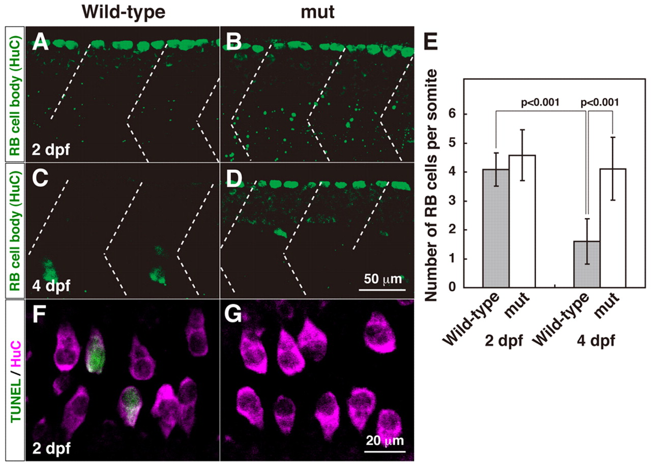
Fig. 2 Programmed cell death of RB neurons is diminished in mi310 mutants. (A) In a lateral view of a wild-type embryo at 2 dpf, anti-HuC labeled large cell bodies of RB neurons (green). Dashed lines represent somite boundaries. (B) A mutant embryo displayed a comparable number of HuC-positive cells at 2 dpf. (C) In wild type, RB neurons were eliminated by 4 dpf. (D) HuC-positive RB cells were observed in mutant larva at 4 dpf. (E) The number of RB neurons is significantly reduced between 2 and 4 dpf in wild type but not in mutants. (F) In a dorsal view of a wild-type embryo, some HuC-positive neurons (magenta) were also positive for TUNEL (green) at 2 dpf. (G) HuC-TUNEL double-positive neurons were rarely observed in mutants.