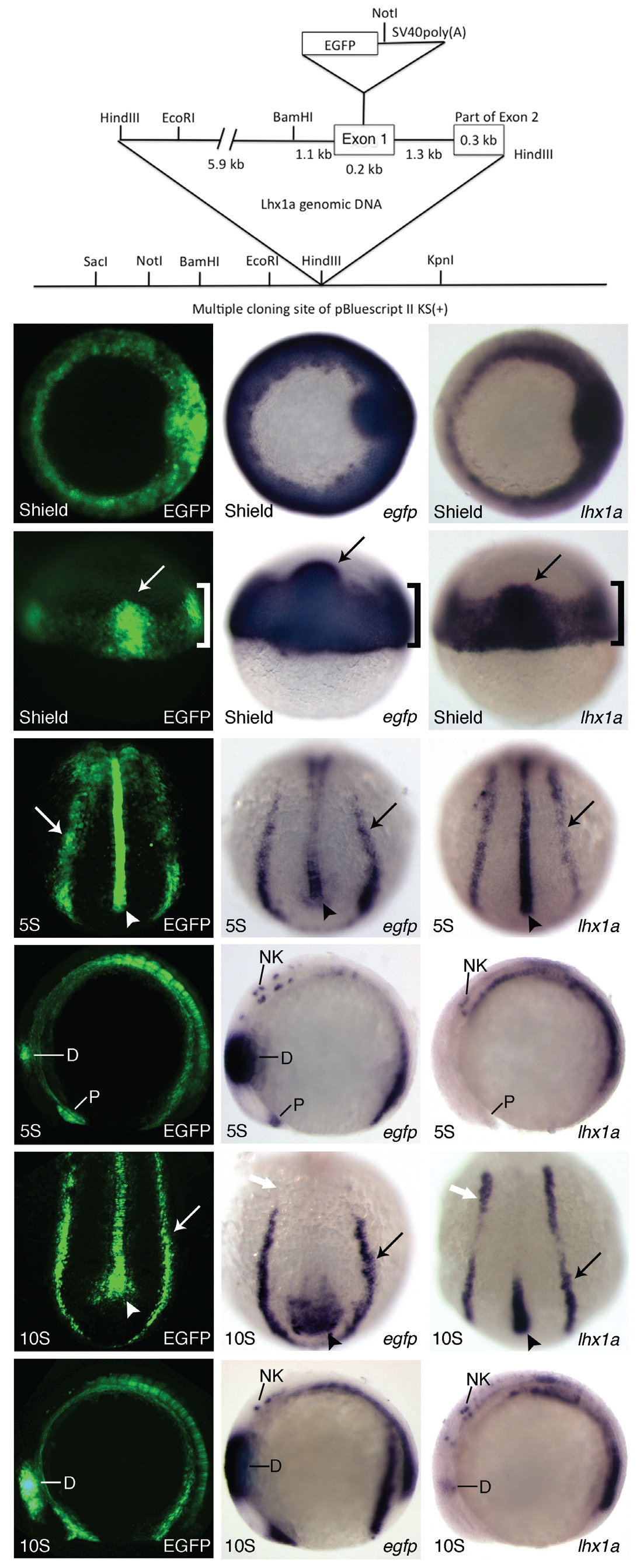
IMAGE

Fig. 1

ID
ZDB-IMAGE-100322-19
Genes
Source
Figures for Swanhart et al., 2010
Image
Figure Caption

Fig. 1 Construct design and early Tg(lhx1a:EGFP)pt303 expression.
(A)
Schematic of lhx1a:EGFP construct. (B-S) Tg(lhx1a:EGFP)pt303 embryos. (B-G) Tg(lhx1a:EGFP)pt303 expression. In situ hybridization for egfp. (N-S) In situ hybridization for lhx1a. (B,H,N) Shield stage embryos, animal view. (C,I,O) Shield stage embryos, dorsal view. Arrow marks the shield and bracket denotes the marginal cells. (D,J,P) 5-somite stage embryos, dorsal-posterior view. Arrow points to intermediate mesoderm and arrowhead marks the notochord. (E,K,Q) 5-somite stage embryos, lateral view. Anterior is to the left. (F,L,R) 10-somite stage embryos, dorsal-posterior view. Arrow points to the intermediate mesoderm and arrowhead marks the notochord. White arrow denotes anterior expression domain of lhx1a in the intermediate mesoderm. (G,M,S) 10- somite stage embryos, lateral view. Anterior is to the left. Abbreviations: D, diencephalon; NK, neural keel; P, polster.

Figure Data
Acknowledgments
This image is the copyrighted work of the attributed author or publisher, and ZFIN has permission only to display this image to its users. Additional permissions should be obtained from the applicable author or publisher of the image. Full text @ Int. J. Dev. Biol.