
Fig. 7
- ID
- ZDB-IMAGE-100223-40
- Publication
- Raeker et al., 2010 - Targeted deletion of the zebrafish obscurin A RhoGEF domain affects heart, skeletal muscle and brain development
- All Figures
- Figures for Raeker et al., 2010
|
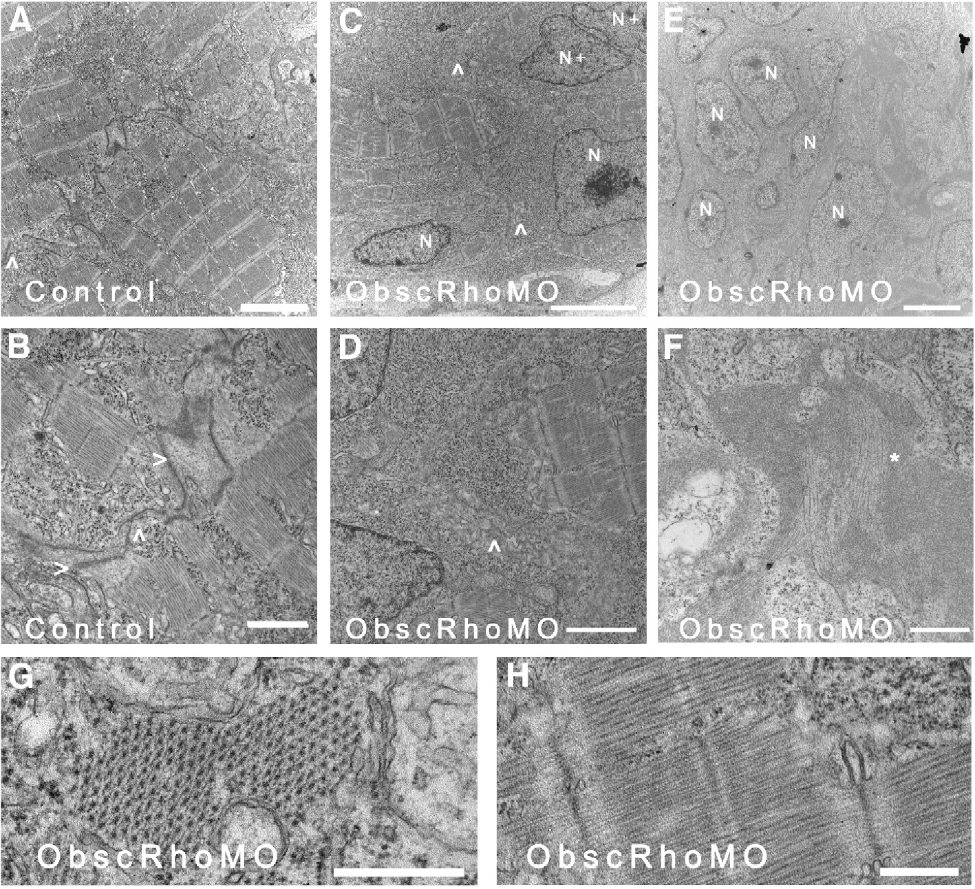
Fig. 7 Ultrastructural analysis of cardiac myocytes at 72 hpf. (A) Cardiac myocytes in control embryos have formed intercalated disks (^) and filled the sarcoplasm with mature myofibrils. (B) Higher magnification of an intercalated disk (ID) from a control embryo demonstrates adherens (>) and gap (^) junctions. (C, D) In contrast, cardiac myocytes from mildly affected obscurin A RhoGEF morphant embryos have reduced myofibrillar content [compare the nuclear (N) area to the myofibrillar area within each myocyte] and very poorly defined regions of cell–cell (^) contact with no identifiable IDs. Examples of recent nuclear division (N+) were more frequent in the obscurin RhoGEF morphant embryos. (E, F) In more severely affected embryos, cardiac myocytes appear markedly abnormal with loosely arranged contractile filaments (F: ∗). (G, H) The mature cardiac myofibrils that did form in the obscurin A RhoGEF morphant embryos were not significantly different than control with a normal appearing lattice of thick filaments on cross-section (G) and laterally aligned Z disks and M bands on longitudinal section (H). Scale bars are 4 μm (A–C), 2 μm (D, E), 1 μm (F) and 0.5 μm (G, H).
Reprinted from Developmental Biology, 337(2), Raeker, M.O., Bieniek, A.N., Ryan, A.S., Tsai, H.J., Zahn, K.M., and Russell, M.W., Targeted deletion of the zebrafish obscurin A RhoGEF domain affects heart, skeletal muscle and brain development, 432-443, Copyright (2010) with permission from Elsevier. Full text @ Dev. Biol.