
Fig. 4
- ID
- ZDB-IMAGE-100223-37
- Publication
- Raeker et al., 2010 - Targeted deletion of the zebrafish obscurin A RhoGEF domain affects heart, skeletal muscle and brain development
- All Figures
- Figures for Raeker et al., 2010
|
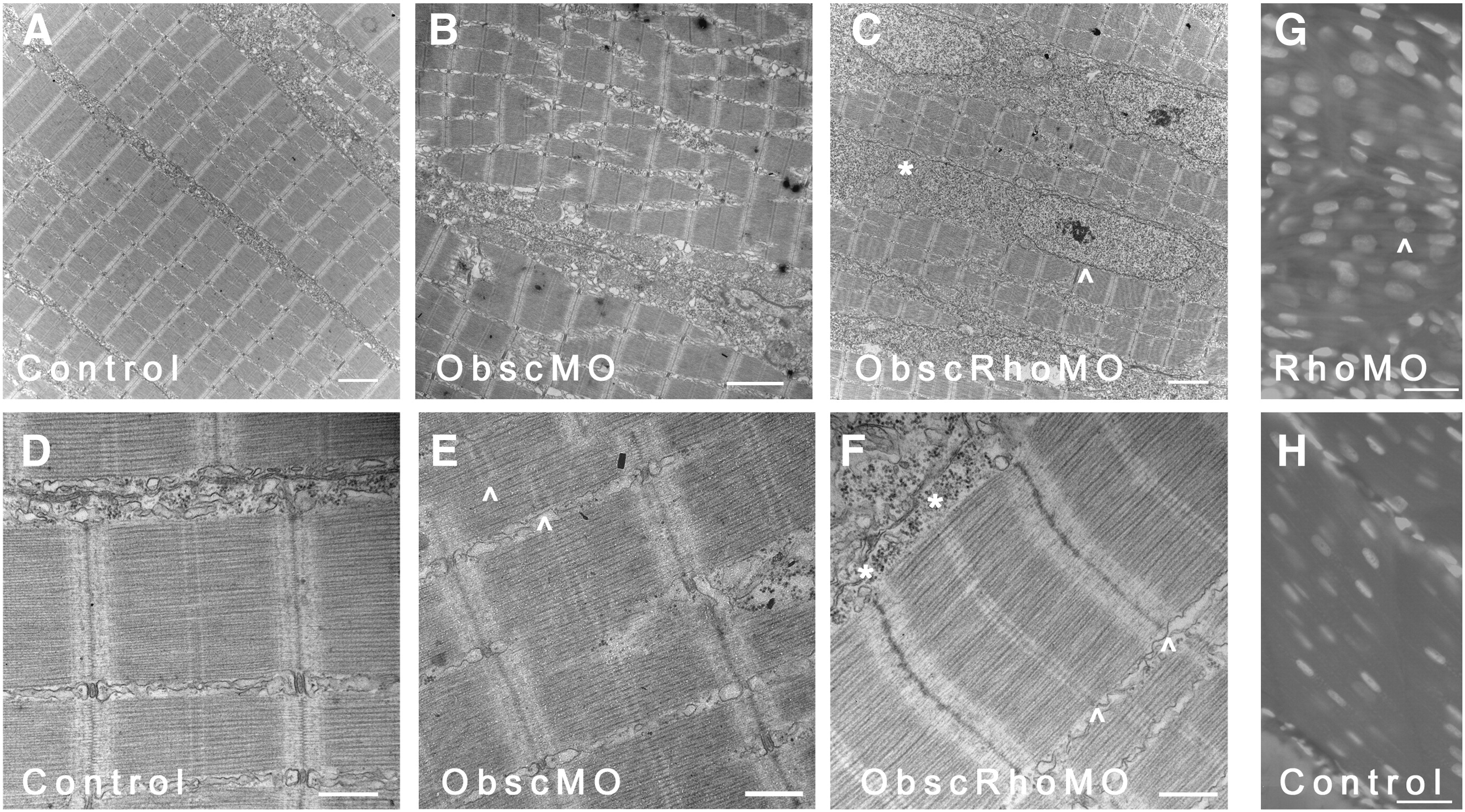
Fig. 4 Skeletal muscle phenotype in obscurin A RhoGEF morphant embryos at 72 hpf. Ultrastructural analysis was performed at 72 hpf on control, obscurin A (ObscMO) and obscurin A RhoGEF (ObscRhoMO) morphant embryos. In control embryos, the myofibrils are aligned in register across the sarcoplasm (A) with well defined M bands and finely “stitched” Z disks (D). In the obscurin morphant embryos (B) there is marked disturbance of myofibril organization with areas of misalignment of the M bands of adjacent myofibrils (E: ^). By comparison, the obscurin A RhoGEF morphants display myofibril organization (C) similar to control embryos but reduced myofibrillar content with more sarcoplasm devoid of myofibrils and more rounded nuclei (C, G: ^). There were also mild sarcomeric abnormalities including, infrequently, an indistinct appearance of the Z disk and absence of the M band (F: ∗) which could occur even within myofibrils demonstrating mature Z disks and M bands (F: ^). Note that in skeletal myocytes stained with DAPI, the nuclei in the obscurin A RhoGEF morphants (RhoMO) are closer together and more rounded (G) than in controls (H) consistent with reduced myofibril volume. Scale bars 2 μm (A–C), 0.5 μm (D–F) and 10 μm (G, H).
Reprinted from Developmental Biology, 337(2), Raeker, M.O., Bieniek, A.N., Ryan, A.S., Tsai, H.J., Zahn, K.M., and Russell, M.W., Targeted deletion of the zebrafish obscurin A RhoGEF domain affects heart, skeletal muscle and brain development, 432-443, Copyright (2010) with permission from Elsevier. Full text @ Dev. Biol.