
Fig. 5
- ID
- ZDB-IMAGE-091214-5
- Genes
- Antibodies
- Publication
- Bibliowicz et al., 2009 - Expanded progenitor populations, vitreo-retinal abnormalities, and Muller glial reactivity in the zebrafish leprechaun/patched2 retina
- All Figures
- Figures for Bibliowicz et al., 2009
|
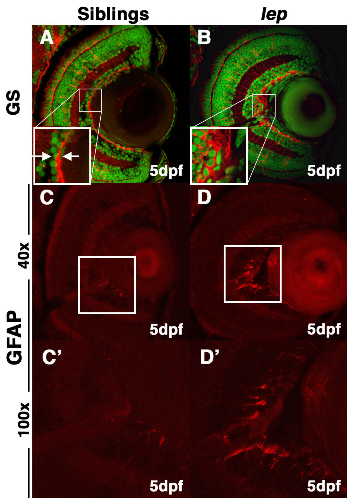
Fig. 5 lep/ptc2 mutants display Müller glial reactivity and morphological abnormalities in the ILM. Immunohistochemical analysis using glutamine synthetase (GS) antibody, which marks differentiated Müller glia and their endfeet at the ILM, highlights disruptions in the ILM. (A) In sibling retinas, the ILM is tight and continuous (inset). (B) In lep/ptc2 retinas the ILM is discontinuous and Müller glial endfeet do not terminate properly at the ILM (inset). Glial fibrillary acidic protein (GFAP) antibody staining reveals elevated immuno-reactivity in the inner retina, adjacent to the optic nerve of lep/ptc2 (D and arrows in D′) mutant retinas as compared to siblings (C, C′). Approximately 40% of analyzed mutants displayed significant ILM disruptions and elevated GFAP levels.