
Fig. 2
- ID
- ZDB-IMAGE-090826-21
- Genes
- Publication
- Seipold et al., 2009 - Non-SMC condensin I complex proteins control chromosome segregation and survival of proliferating cells in the zebrafish neural retina
- All Figures
- Figures for Seipold et al., 2009
|
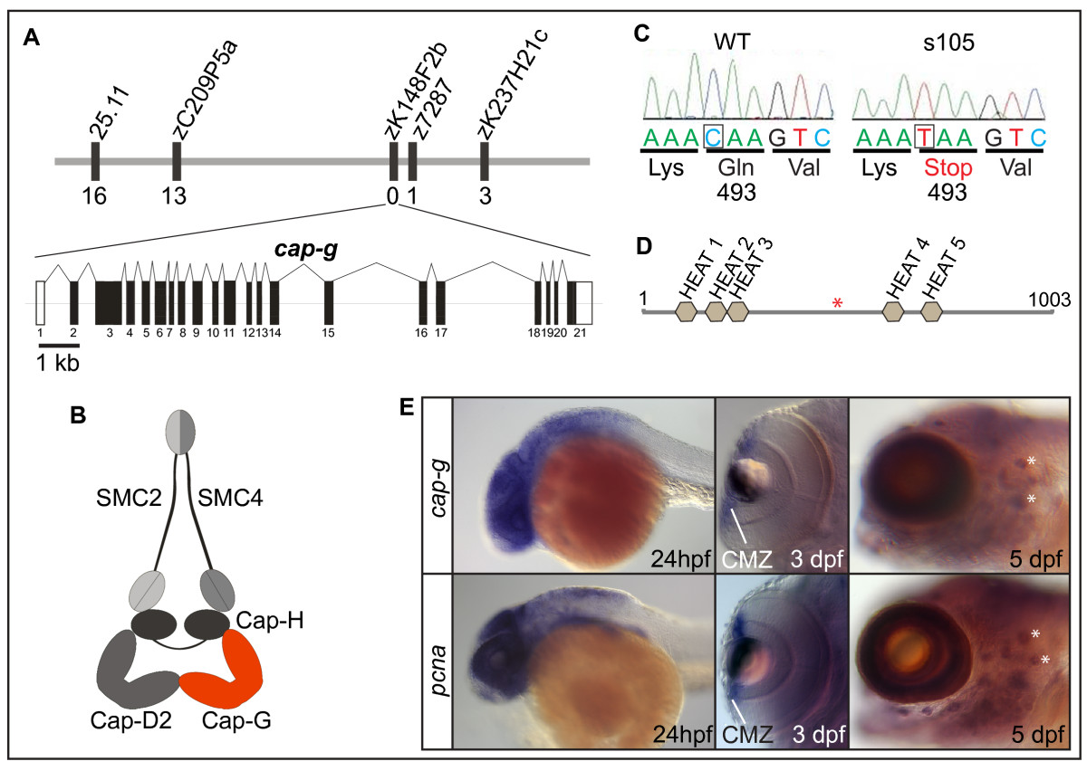
Fig. 2 The zebrafish cap-g gene is mutated in s105. (A) Representation of the genetic map of the cap-g locus on linkage group 1 (LG1) and exon/intron structure of the cap-g transcript. Some of the markers utilized for cloning the mutation are indicated and the number of recombinants among 2982 meioses is indicated below each marker. (B) Schematic model of the associated condensin I complex. (C) Comparison of sequence data for wild-type and s105 mutant alleles. The s105 mutation generates a premature stop codon. (D) Schematic diagram of the Cap-G protein which contains several predicted HEAT domains. The s105 mutation generates a premature stop codon (red asterisk) that truncates more than half of the protein. (E) Comparison of cap-g expression with that of pcna by whole-mount in situ hybridization. Overlapping expression with pcna within the brain, the CMZ of the retina, which contains retinal stem cells, and within neuromasts of the lateral line organ (white asterisks) indicates that cap-g is required within proliferative cells.