
Fig. 5
- ID
- ZDB-IMAGE-090716-6
- Genes
- Publication
- Paridaen et al., 2009 - Apc1 is required for maintenance of local brain organizers and dorsal midbrain survival
- All Figures
- Figures for Paridaen et al., 2009
|
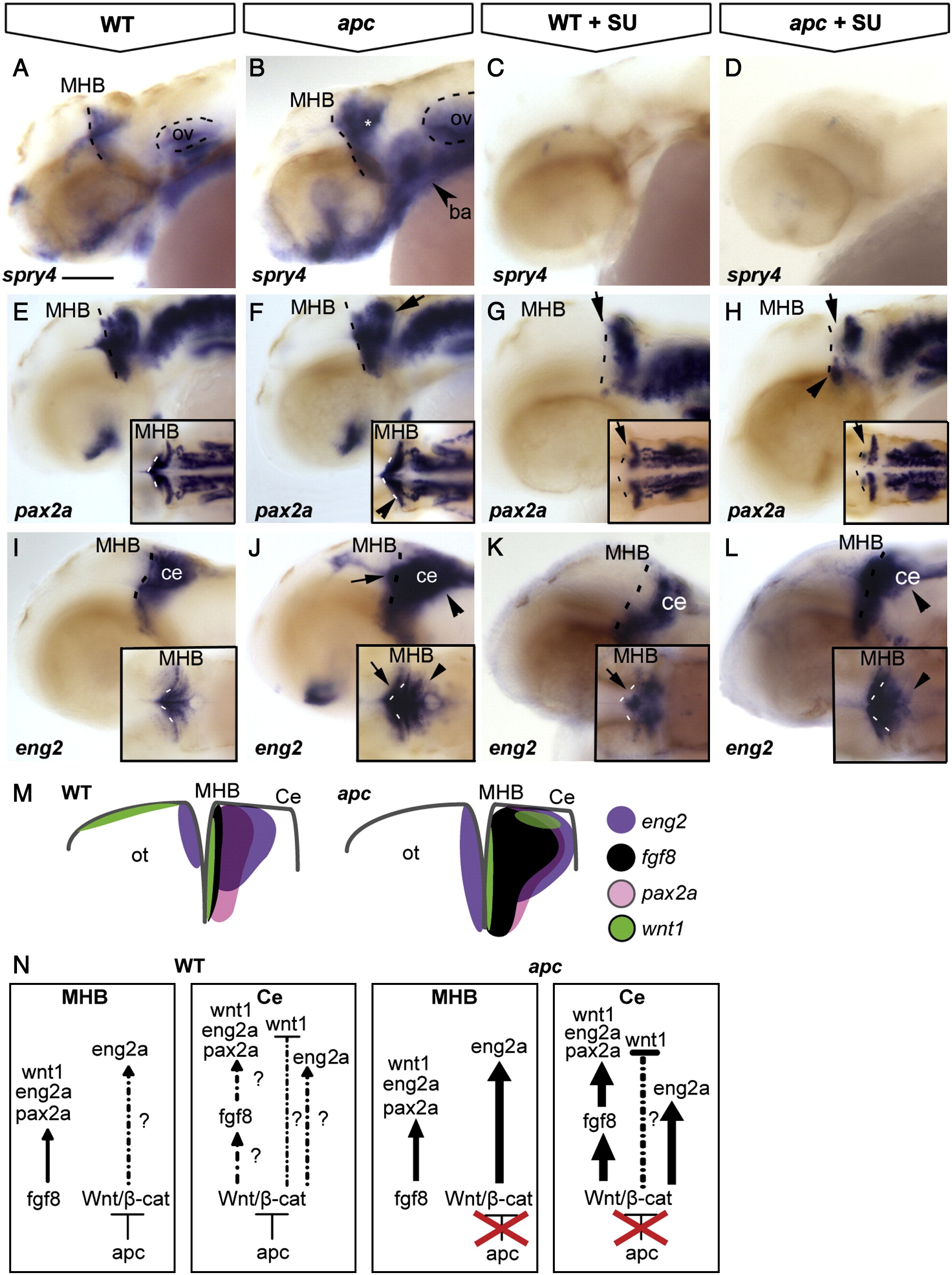
Fig. 5 The contribution of Wnt/β-catenin and Fgf8 signalling to maintenance of the IsO. Lateral view, anterior to the left. Embryos at 42 hpf. (A, B) The Fgf8 target spry4 is expanded in the MHB, cerebellum (asterisk) and branchial arches (arrowhead) of apc mutants. (C, D) Upon treatment with 8 and 25 μM SU5402, spry4 is lost from both wild-type and mutant embryos. (E–H) pax2a is expanded laterally at the MHB (arrowhead in inset F) and caudally into the cerebellum of apc mutants (arrow) as compared to wild-types. (G, H) After treatment with 25 μM SU5402, pax2a is abolished from the MHB (arrow) of wild-type embryos (G) and in apc mutants (H). pax2a expression in the ventral MHB of apc mutants appeared refractory to Fgf inhibition (arrowhead in panel H), although pax2a expression is absent from the pons in treated apc mutants. (I–L) In wild-type embryos (I, K), SU5402 treatment caused disruption of eng2a expression at the MHB (arrow). However, in apc mutants, eng2a expression persisted in the MHB upon SU5402 treatment (L). In contrast, eng2a expression in the cerebellum is slightly downregulated (arrowhead in panel L) as compared to untreated mutants (arrowhead in panel J;). Dashed line indicates the MHB. Insets - dorsal view. (M) Schematic view showing expression of regulatory genes in the isthmic region. (N) Model showing regulation of IsO genes by Fgf8 and Wnt/β-catenin. In wild-type embryos, Apc1 expression in the cerebellum mediates restriction of Fgf8 expression by limiting Wnt/β-catenin signalling and thereby preventing caudal expansion of the isthmic markers pax2a, wnt1 and eng2a that are regulated by Fgf8. In apc mutants, the restriction of Wnt/β-catenin signalling is lifted, and overactivated Wnt/β-catenin signalling causes direct caudal expansion into the cerebellum and MHB of eng2a and indirect expansion of pax2a through ectopic Fgf8. wnt1 expression in the dorsal midbrain is not regulated by Fgf8 and hence remains absent in apc mutants (data not shown). ba, branchial arches; ce, cerebellum; MHB, mid–hindbrain boundary; ot, optic tectum; ov, otic vesicle. Scale bar 125 μm.
Reprinted from Developmental Biology, 331(2), Paridaen, J.T., Danesin, C., Elas, A.T., van de Water, S., Houart, C., and Zivkovic, D., Apc1 is required for maintenance of local brain organizers and dorsal midbrain survival, 101-112, Copyright (2009) with permission from Elsevier. Full text @ Dev. Biol.