
Fig. 7
- ID
- ZDB-IMAGE-090710-38
- Publication
- Lunt et al., 2009 - Zebrafish ift57, ift88, and ift172 intraflagellar transport mutants disrupt cilia but do not affect hedgehog signaling
- All Figures
- Figures for Lunt et al., 2009
|
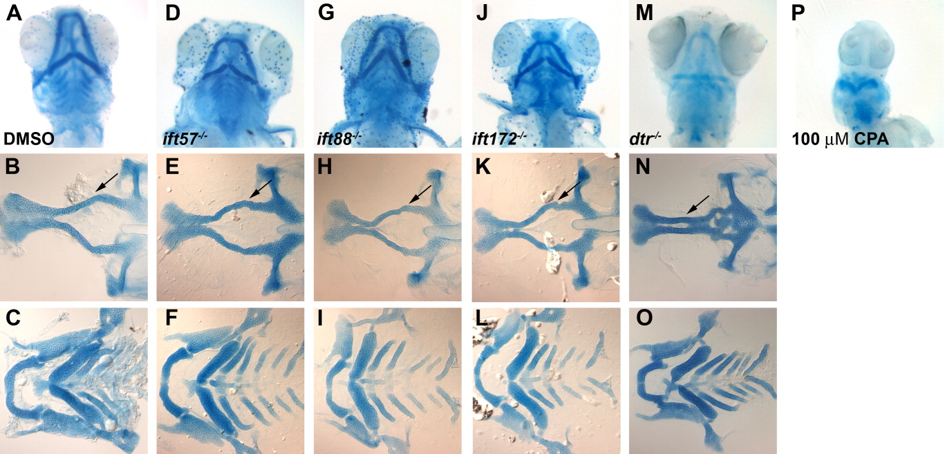
Fig. 7 Alcian blue staining of jaw and brachial arch structure at 5 dpf. A-C: Wild type embryos raised in DMSO vehicle exhibit normal craniofacial head skeleton. D-L: IFT mutants displayed no reduction or loss of the major head structures and no fusion of the trabeculae. M-O: detourgli1 mutants showed significant narrowing of ethmoid plate (black arrow). P: Embryos treated with 100 μM cyclopamine lack all major head cartilage. The top row shows whole-mount images viewed from the ventral side. The middle row shows flat-mount images of dissected dorsal neurocranium structures. The bottom row shows flat-mounted images of dissected ventral structures to show brachial arches.