
|
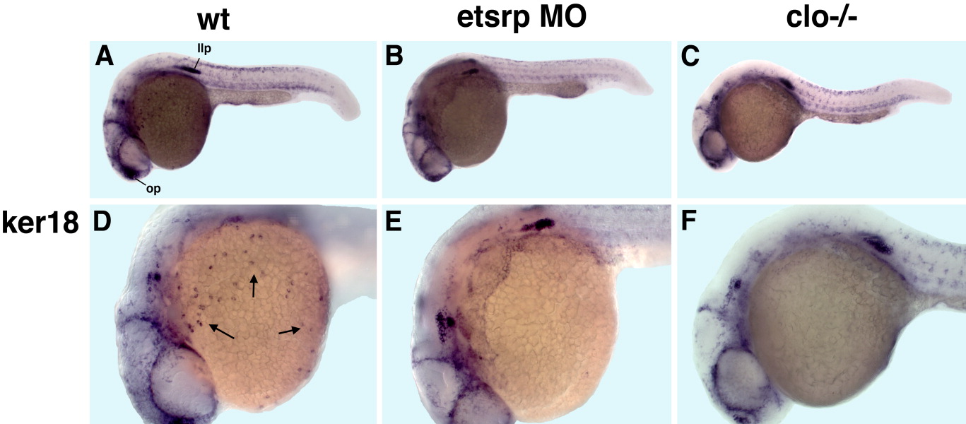
Fig. 6 A-F: Expression of keratin18 (ker18) in control wild-type embryos (A,D), etsrp morphants (B,E), and homozygous cloche mutant embryos (C,F) at 24 hours postfertilization (hpf). Lateral view, anterior is to the left. A-C: Ker18 expression in epidermal cells, neural cells including the lateral line primordia and olfactory placodes is not affected in etsrp morphants and clo mutants. Note that ker18 expression is apparent within the superficial epidermal tissue and no vascular-specific staining is observed at these stages. D-F: However, multiple ker18-expressing cells that are located on top of the yolk and apparently correspond to the myeloid cells (arrows, D), are absent in etsrp morphants and clo mutants (D-F).