
Fig. 6
- ID
- ZDB-IMAGE-090504-14
- Genes
- Antibodies
- Publication
- Zeng et al., 2009 - Phospholipase D1 is required for angiogenesis of intersegmental blood vessels in zebrafish
- All Figures
- Figures for Zeng et al., 2009
|
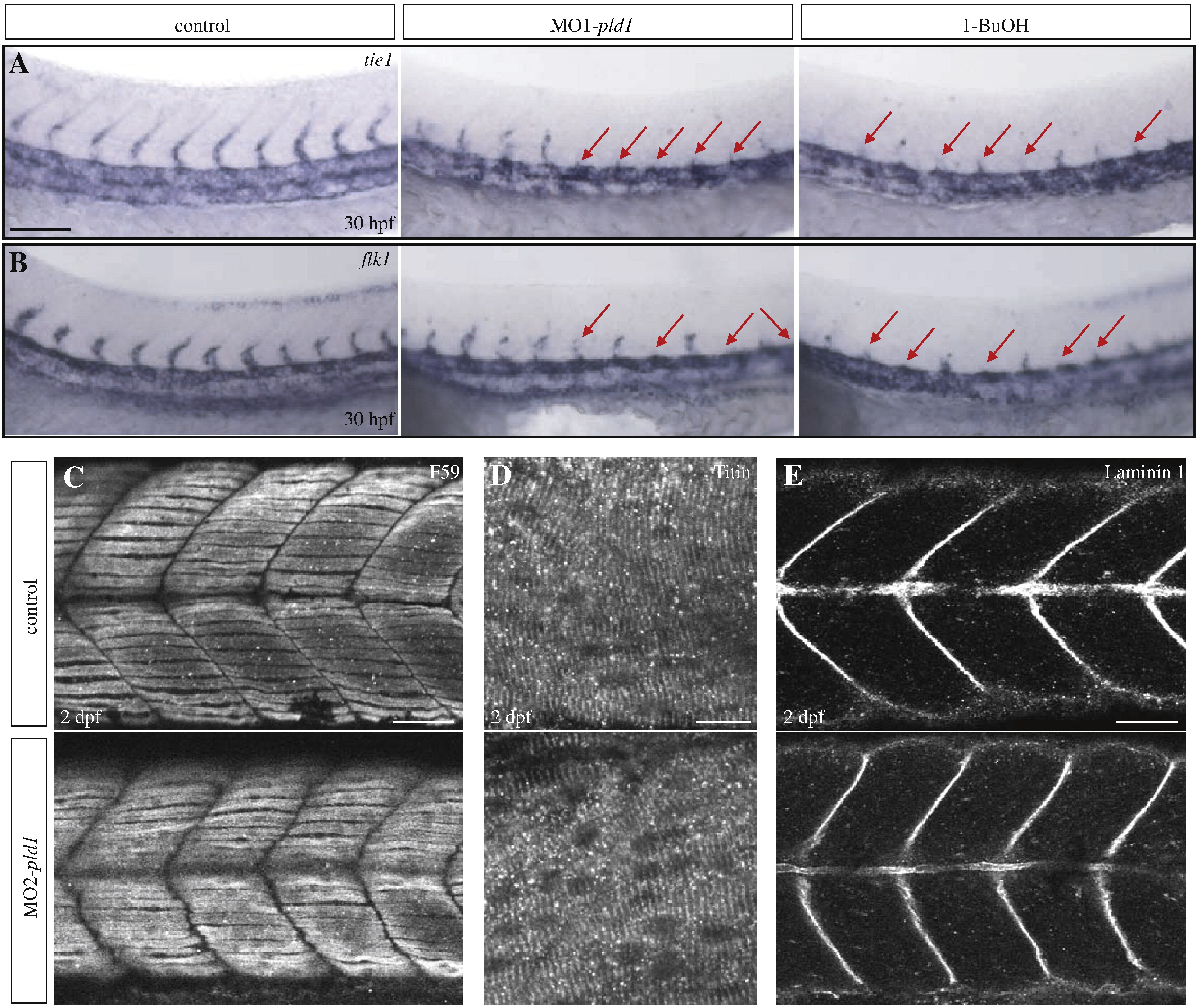
Fig. 6 Effect of Pld1 deficiency on ISV and surrounding tissue development. (A, B) Loss of Pld1 function impairs ISV development. Whole mount in situ hybridization showing expression of tie1 (A) and flk1 (B) at 30 hpf in the trunk region of uninjected control embryos, and embryos either injected with MO1-pld1 or treated with 1-butanol (1-BuOH). Red arrows indicate stunted ISVs. (C–E) Somite muscle and notochord development is relatively normal in embryos with disrupted Pld1 signaling. Representative confocal images of whole-mount immunohistochemistry show the morphology of somites, slow muscle and notochord in MO2-pld1 injected embryos at 2 dpf. Lateral view, anterior to the left. (C) F59 antibody was used to stain slow muscle Myosin. (D) Titin antibody was used to label the organization of muscle cell “Z-discs”. (E) Laminin immunoreactivity at the myosepta and the notochord region in control sibling and pld1 morphants embryos. Scale bar represents 10 μm (D), 20 μm (C, E) and 40 μm (A).
Reprinted from Developmental Biology, 328(2), Zeng, X.X., Zheng, X., Xiang, Y., Cho, H.P., Jessen, J.R., Zhong, T.P., Solnica-Krezel, L., and Brown, H.A., Phospholipase D1 is required for angiogenesis of intersegmental blood vessels in zebrafish, 363-376, Copyright (2009) with permission from Elsevier. Full text @ Dev. Biol.