
Fig. 1
- ID
- ZDB-IMAGE-090127-8
- Genes
- Antibodies
- Publication
- Noël et al., 2008 - Organ-specific requirements for Hdac1 in liver and pancreas formation
- All Figures
- Figures for Noël et al., 2008
|
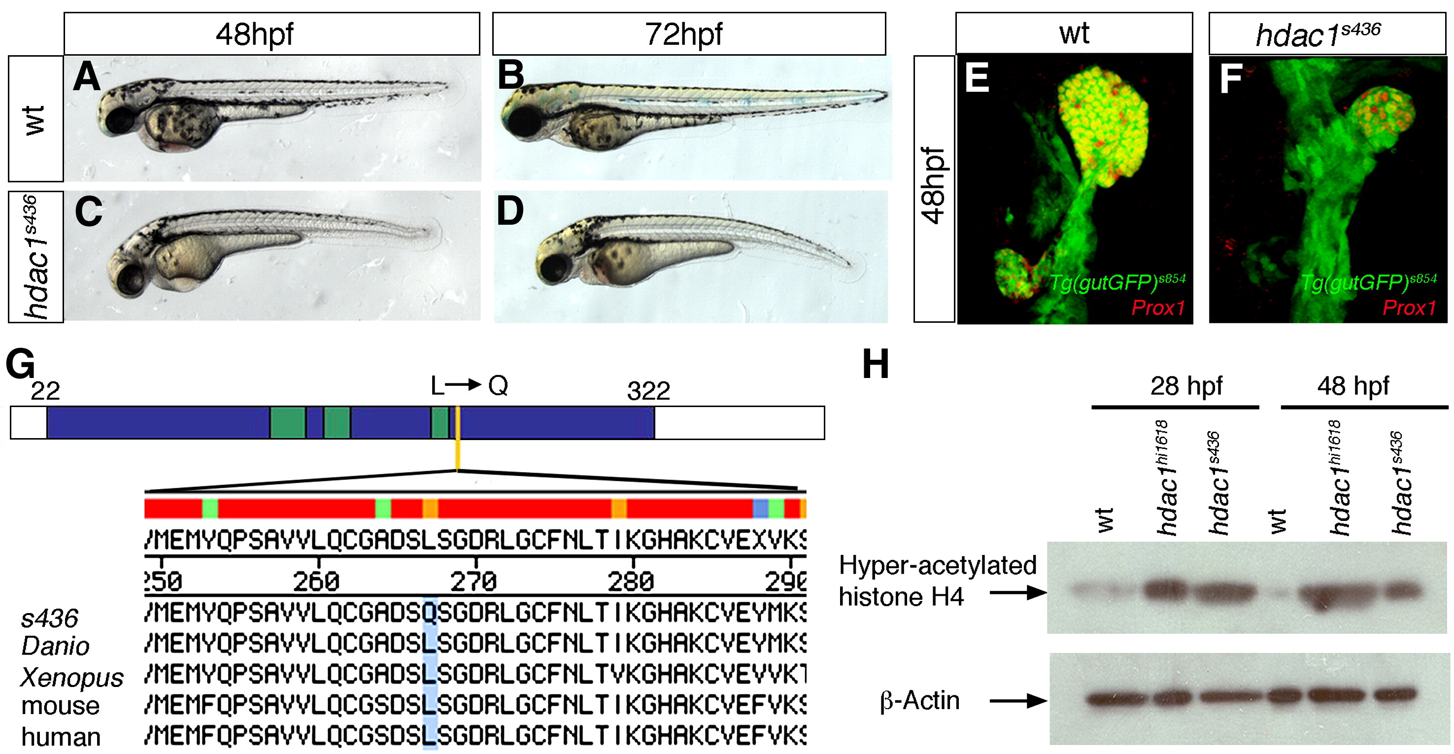
Fig. 1 Mutant line s463 encodes a novel hdac1 allele. Lateral brightfield views of sibling (A, B) and hdac1s436 mutant (C, D) embryos at 48 hpf and 72 hpf, anterior to the left. (E, F) Projections of confocal stacks showing ventral views of Tg(gutGFP)s854, anterior to the top. (E) Wild type embryo at 48 hpf, expressing Prox1 (red) in the liver and exocrine pancreas. (F) s436 mutant exhibits a reduced liver and absent exocrine pancreas at 48 hpf. (G) s436 encodes a novel allele of Hdac1. A T to A base pair change at position 800 results in a leucine to glutamine transition at position 267. Schematic representation of the predicted zebrafish Hdac1 derived from alignment of vertebrate sequences; catalytic domain is depicted in blue, HDAC superfamily signature sequences in green, s436 lesion in yellow. Protein alignments with amino acid transition shown in blue. (H) Western blot showing increased levels of acetylated histone H4 in both hdac1hi1618 and hdac1s436 mutant embryos at 28 hpf and 48 hpf. β-actin levels were used as loading control.
Reprinted from Developmental Biology, 322(2), Noël, E.S., Casal-Sueiro, A., Busch-Nentwich, E., Verkade, H., Dong, P.D., Stemple, D.L., and Ober, E.A., Organ-specific requirements for Hdac1 in liver and pancreas formation, 237-250, Copyright (2008) with permission from Elsevier. Full text @ Dev. Biol.