
Fig. 1
- ID
- ZDB-IMAGE-081117-1
- Genes
- Publication
- Aman et al., 2008 - Wnt/beta-catenin and Fgf signaling control collective cell migration by restricting chemokine receptor expression
- All Figures
- Figures for Aman et al., 2008
|
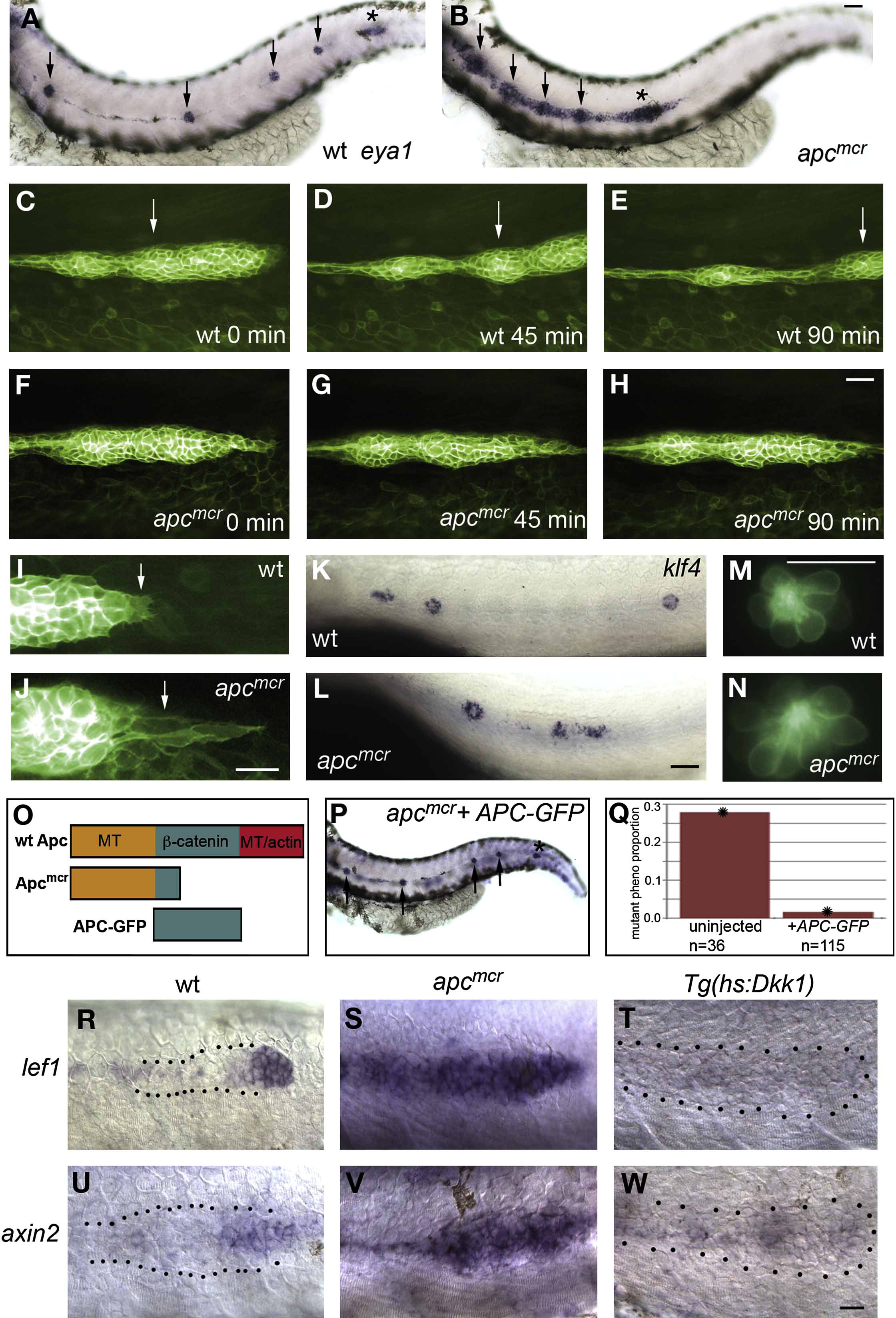
Fig. 1 Constitutive Activation of Wnt/β-Catenin Signaling Disrupts Primordium Migration, but Not Cell Type Specification
Anterior is oriented toward the left in all figures.
(A and B) In situ hybridization with the lateral line marker eya1 of 36 hpf WT and apcmcr mutant embryos. (A) In WT embryos, the primordium (asterisk) has deposited neuromasts (arrows) and almost reached the tail tip. (B) In mutant embryos, the primordium (asterisk) stopped migrating before reaching the tail.
(C–E) Still images of a 90 min time-lapse recording of a normally migrating lateral line primordium in a Tg(Cldnb:lynGFP) embryo. (C) 34 hpf, time point 0 min. (D) After 45 min. (E) After 90 min. The trailing zone of the primordium (arrow) has almost left the field of view.
(F–H) Still images of a 90 min time-lapse recording of a Tg(Cldnb:lynGFP);apcmcr mutant primordium. (F) 34 hpf, time point 0 min. (G) After 45 min. (H) After 90 min. The primordium is not moving tailward.
(I and J) Higher-magnification images of tip cells (arrows) in migrating (I) Tg(Cldnb:lynGFP) and (J) Tg(Cldnb:lynGFP);apcmcr mutant primordia.
(K–N) (K and L) Support and (M and N) hair cells are specified in apcmcr mutant embryos. klf4 in situ labels supporting cells in 36 hpf (K) WT and (L) apcmcr mutants. Hair cells in (M) Tg(Brn3c:GAP43-GFP)s356t and (N) Tg(Brn3c:GAP43-GFP)s356t;apcmcr 72 hpf mutant embryos.
(O) Simplified protein structure of WT Apc, Apcmcr, and APC-GFP.
(P) An apcmcr embryo that was rescued by APC-GFP injection as revealed by eya1 in situ. Arrows indicate neuromasts; an asterisk labels the primordium.
(Q) Quantification of the rescue effect. Rescue was found to be significant by a test of difference in population proportions (p = 2.3 x 10⁻⁷).
(R–W) (R and U) Wnt target genes lef1 and axin2 are expressed in the tip of 36 hpf WT primordia. (S and V) In apcmcr mutant embryos, lef1 and axin2 are expressed in the entire primordium and deposited cells. (T and W) Tg(hs:Dkk1) embryos, heat shocked at 20 hpf, express no lef1 or axin2 by 4 hr post-heat shock.
Scale bars in (A)–(L) and (R)–(W) are equal to 40 μM. Scale bars in (M) and (N) are equal to 20 μM.
Reprinted from Developmental Cell, 15(5), Aman, A., and Piotrowski, T., Wnt/beta-catenin and Fgf signaling control collective cell migration by restricting chemokine receptor expression, 749-761, Copyright (2008) with permission from Elsevier. Full text @ Dev. Cell