
Fig. 6
- ID
- ZDB-IMAGE-081021-156
- Genes
- Antibodies
- Publication
- Grosskurth et al., 2008 - Emergence of Xin demarcates a key innovation in heart evolution
- All Figures
- Figures for Grosskurth et al., 2008
|
Fig. 6
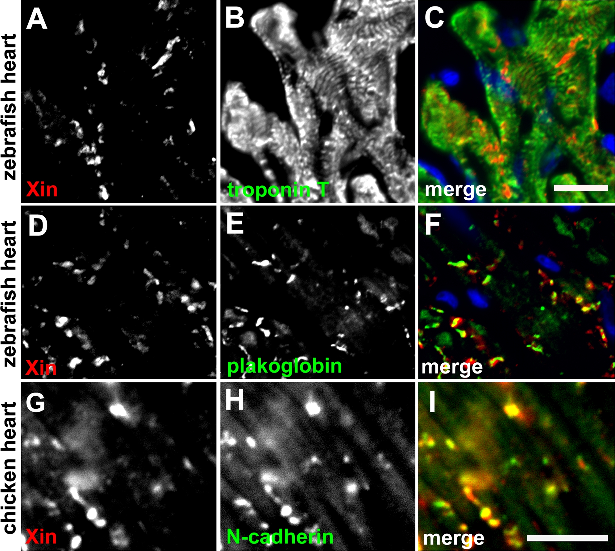
Immunofluorescence microscopy of zebrafish and chicken heart sections.
(A–F) Double-label indirect immunofluorescence was performed on frozen sections of zebrafish heart with monoclonal anti-troponin T or anti-plakoglobin (green, visualized by fluorescein-conjugated 2nd antibody) and polyclonal anti-mXin (red, visualized by rhodamine-conjugated 2nd antibody). Before mounting, the sections were treated with DAPI to stain nuclei (blue). Scale bar: 10 μm. (G–I) Double-label indirect immunofluorescence was performed on chicken heart section with monoclonal anti-N-cadherin (green) and polyclonal anti-mXin antibody (red). Scale bar: 10 μm.