
Fig. 5
- ID
- ZDB-IMAGE-081021-155
- Genes
- Antibodies
- Publication
- Grosskurth et al., 2008 - Emergence of Xin demarcates a key innovation in heart evolution
- All Figures
- Figures for Grosskurth et al., 2008
|
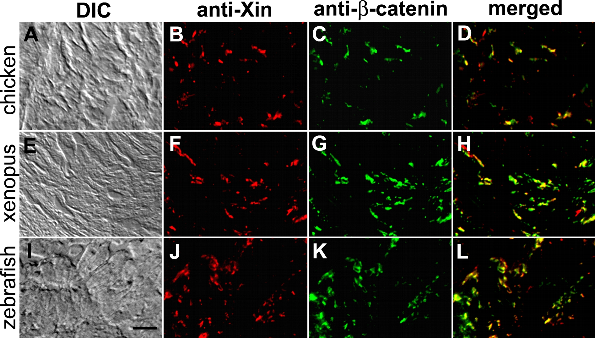
Fig. 5
Co-localization of Xin proteins and β-catenin in chicken, frog and zebrafish hearts.
Double-label indirect immunofluorescence microscopy was performed on frozen sections of chicken (A–D), frog (E–H) and zebrafish (I–L) hearts with mouse monoclonal anti-β-catenin (C, G, K) and rabbit polyclonal U1013 anti-mXin (B, F, J) antibodies, and subsequently with a mixture of rhodamine-conjugated goat anti-rabbit IgG and fluorescein-conjugated goat anti-mouse IgG. Merged images (D, H, L) indicate co-localization of the two proteins. The differential interference-contrast (DIC) images correspond to the fluorescent images shown in respectively. Scale bar: 10 μm.